Computerized Tomography (CT)
Despite the fact that MR has largely superseded CT in defining the anatomy, CT remains a necessary tool for the neurosurgeon in a variety of circumstances. CT is used routinely in emergency settings and in the immediate postoperative period due to its availability, short acquisition time and sensitivity in demonstrating potential bleeding complications. This study is also incredibly sensitive in assessing for hydrocephalus and ventricular anatomy. For tumors of the pituitary and skull base, CT provides a superior view of intricate and delicate bony anatomy of the paranasal sinuses and inner ear. CT also gives a detailed view of bony spinal anatomy and the adequacy of positioning of spinal implants.
CT Angiography (CTA)
This diagnostic and non-invasive test is exceedingly sensitive in assessing the cerebral vascular anatomy and detecting the presence of cerebral aneurysm. It has a variety of applications including determining the presence of vasospasm in subarachnoid hemorrhage and is the study of choice for intra-operative navigation in endonasal endoscopic surgery.

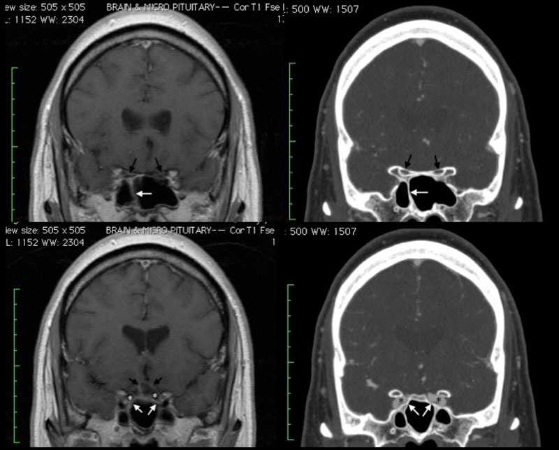
Figure 1
These images highlight some of the differences between CT (right) and MR (left) imaging. Coronal views of the skull base demonstrate details of the bony anatomy of the sinuses (white arrow) as well as the bony canals transmitting the optic nerve (black arrows) with greater clarity than on MR. CT angiography, furthermore, outlines with precision the relationship of arteries (asterisk) to the overlying bony structures. Note, however, the detail of brain anatomy seen on MR that is not well visualized on CT.

