We are a diverse team of experts in advanced neuro-oncology techniques and approaches. Our subspecialized team of neuro-oncologists, radiation oncologists, medical neuro-oncologists, neuroradiologists, and neurologists partner closely on every case to develop a highly individualized treatment plan that is right for you.
What are intraventricular tumors?
Ventricles are cavities within the brain that are filled with a clear, protective liquid called cerebrospinal fluid. When brain tumors occur within these cavities, they are known as intraventricular tumors, of which there are several types:
- Astrocytomas: tumors that originate from supporting cells in the brain
- Meningiomas: tumors that form in the brain’s protective covering
- Ependymomas: tumors that form within the lining of the ventricles
- Colloid cysts & Craniopharyngiomas: tumors that are formed from developmental cells
What are the symptoms of an intraventricular tumor?
Early symptoms include headaches, nausea, vomiting, vision changes, and cognitive difficulties. As many of these symptoms are prevalent across a variety of illnesses and conditions, it is important to seek medical attention early if you experience several of these symptoms together.
Because of their location, these tumors often block the flow of cerebrospinal fluid, causing a build-up known as obstructive hydrocephalus. In hydrocephalus, the additional fluid increases pressure on surrounding brain tissue and causes a variety of symptoms depending on location that over time become life-threatening:
- Headache
- Nausea
- Mental status deterioration
- Sight disruption
- Seizures
- Weakness, tingling, numbness in limbs
- Difficulty speaking
- Mood changes
- Memory loss
- Eventually, death
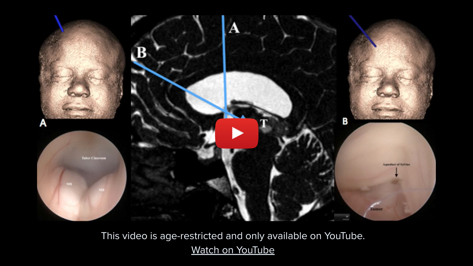
What is an endoscopic biopsy of the brain?
To help determine the best course of treatment for an intraventricular tumor, a neurosurgeon would begin by taking a biopsy, a small sample of tissue, from the tumor using a technique called neuroendoscopy. This is a technique in which the neurosurgeon uses a long, flexible, lighted tube with a high-resolution camera called an endoscope. The endoscope is inserted through the nasal cavity, making it a minimally invasive method to examine the brain.
Specialized tools are then inserted through the tube, which allow the surgeon to take tissue samples from or around the ventricles for testing. This is called an endoscopic biopsy.
What is the difference between a biopsy and a resection?
A biopsy is useful for diagnosis, while a resection is used for treatment. During a biopsy, a small tissue sample, or samples, will be collected to examine and diagnose, while a resection is a surgery used to remove a large portion – or all – of a tumor. An endoscopic resection would be a recommended course of a treatment if the biopsy of an intraventricular tumor came back as cancerous.
What is neuroendoscopy for an intraventricular brain tumor?
Neuroendoscopy is a minimally invasive endoscopic surgery that allows a surgeon to diagnose and treat intraventricular brain tumors. The procedure involves using a tool called an endoscope, which is a long tube that can reach the brain and allows the surgeon to see the affected area. The surgeon then works through the endoscope to extract and remove the tumor through the nasal passageway. This is called an endoscopic excision.
Neuroendoscopy is typically more successful for smaller cyst-like tumors. Larger, solid tumors typically require more invasive, technically challenging, traditional forms of surgery like a craniotomy, which involves an incision in the scalp to remove a section of the skull to reach the tumor. However, when these larger, solid tumors are difficult to reach with a craniotomy, endoscopic surgery may be recommended.
What is recovery from a neuroendoscopy like?
A neuroendoscopy to perform endoscopic resection offers several benefits when compared to more conventional surgical approaches. The patient experiences faster recovery time, a reduced risk of complications, and minimal scarring. Typically, the patient will remain in the hospital for monitoring 1-3 days following the procedure, and will usually have a CT scan or MRI to check for early complications. Some sensitivity around the nose and head may be experienced, although pain is usually minimal.
Have More Questions about This Condition

New Jersey Brain and Spine Earns #3 National Ranking For Neurosurgical Practices
New Jersey Brain and Spine has once again been recognized among the nation’s elite neurosurgical practices—this time...

Patient Story: Patient Gets Fitness Career Back in Shape Thanks to Dr. Roth
Robin Bray is the owner of FitnessBarre™, a popular local workout studio in Midland Park, New Jersey. The...

NJBS Highlighted for Advanced Tremor Treatment With Hackensack Meridian Health
New Jersey Brain & Spine was recently featured alongside Hackensack Meridian Health, highlighting the impact of...

Patient Story: Ashley’s Recovery After Cervical Disc Herniation
A stroke at birth left Ashley with cerebral palsy and limited mobility on her right side. So when she woke up at 1...

Back on the Competition Track: A Young Athlete’s Recovery After L4–L5 Disc Herniation Surgery
Severe nerve compression brought life to a halt—lumbar microdiscectomy helped him reclaim strength, mobility, and...

Ground-Breaking HiFu Treatment Re-Opens a Door
Innovative non-invasive essential tremor treatment puts car enthusiast back in the driver’s seat Dan lived with...
")
Full Patient Story: Chris’s Recovery After L4–5 Posterior Lumbar Interbody Fusion (PLIF)
How an L4–5 Posterior Lumbar Fusion Gave Chris Feeling in His Leg Again and His Life Back Chris was only in his early...

Back in the Game: Brian’s Recovery After C5–6 Disc Replacement
Neck pain and cervical radiculopathy stole his mobility, his golf swing, and his comfort, an artificial disc...

New Jersey Brain and Spine Launches $2000 Healthcare Scholarship to Support Next Generation of Healthcare Professionals
Investing in the future of healthcare: Annual scholarship open to high school and undergraduate students from or...