What Is Brain Aneurysm Endovascular Coiling?
Brain aneurysm coiling, or endovascular coiling, is a minimally invasive procedure used to treat brain aneurysms, which are weakened areas of a blood vessel that bulge under the pressure of blood flow. To prevent this area from rupturing and bleeding, a neurosurgeon can use this minimally invasive procedure to block blood flow to the aneurysm. The surgeon will navigate a microcatheter (small, flexible tube) through an artery to the aneurysm, where tiny metal coils are expanded to fill the space and promote clotting to prevent further blood flow to the weakened area of the aneurysm.
Case 1: Coiling of Brain Aneurysm
A: An AP view of an aneurysm of the right internal carotid artery. B: Magnified AP view demonstrating an un-ruptured right internal carotid artery aneurysm (arrow). C: Intra-procedural view of the aneurysm during endovascular coiling while the platinum coils are being deployed within the aneurysm. Note that the aneurysm sac fills with contrast suggesting incomplete embolization. The guide catheter and microcatheter are visible within the normal carotid artery and are used to deliver the coils to the aneurysm. D: Complete embolization (blue arrow).

Case 1: Coiling of Brain Aneurysm
Understanding Brain Aneurysms
A brain aneurysm is a balloon-like bulge or weakened area in a blood vessel wall, typically occurring at vessel branching points. These weak spots can grow over time and may eventually rupture, causing a potentially life-threatening brain hemorrhage. Aneurysms often develop silently until they become large or rupture, making preventative treatment critical for at-risk patients.
According to recent statistics:
- Approximately 6.5 million people in the United States (1 in 50 people) have an unruptured brain aneurysm
- About 30,000 people in the United States suffer a brain aneurysm rupture each year
- Ruptured brain aneurysms are fatal in about 40% of cases
- Of those who survive, approximately 66% suffer permanent damage to the brain or nervous system
- Women are more likely than men to develop brain aneurysms at a ratio of 3:2
Common types of brain aneurysms include:
- Saccular (berry) aneurysms — This is the most common type of brain aneurysm typically occurring in the network of arteries at the base of the brain and appears as a round blood-filled sac on one side of the artery.
- Fusiform aneurysms — This type of aneurysm occurs less commonly than saccular aneurysms and can be found in various locations of the brain. It is noted by the way the blood vessel wall bulges out on all sides, like a spindle.
- Dissecting aneurysms — These occur when a tear develops in the inner layer of a blood vessel wall, which allows blood to flow between the layers of the wall, separating them.
When Brain Aneurysm Endovascular Coiling Is Recommended
Brain aneurysm endovascular coiling is typically recommended in the following situations:
- Unruptured aneurysms with high risk of future bleeding
- Recently ruptured aneurysms
- Aneurysms with difficult surgical access
- Patients who may not tolerate open surgery
- Multiple aneurysms requiring treatment
Your neurosurgeon will carefully evaluate factors like aneurysm size, location, shape, and your overall health to determine if coiling is the appropriate treatment option.
The Brain Aneurysm Endoscopic Coiling Procedure
The brain aneurysm coiling procedure involves several precise steps:
- Anesthesia is administered (general anesthesia as mentioned in the video).
- A small incision is made in the groin and a thin catheter (flexible tube) is inserted into the radial or femoral artery, which are major blood vessels that is easily accessible and provides a path to navigate the catheter through the blood vessels toward the aneurysm using X-ray guidance.
- A smaller microcatheter is inserted into the initial catheter to navigate the metal coils to the aneurysm.
- The coils are placed into the aneurysm to block blood flow.
- A small electrical current is passed through to the coil, which causes it to detach and remain inside the aneurysm.
- The catheters are then removed and the incision point closed.
As explained in the physician video, brain aneurysm coiling typically takes 1-3 hours depending on the complexity of the procedure. The goal is to pack the aneurysm approximately 20% to 30% full of metal to cause it to clot and stop blood flow into and out of the aneurysm.
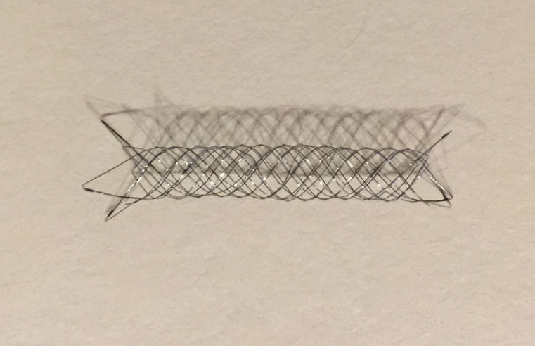
As reviewed in the cerebral angiography section above, a small plastic tube (catheter) is placed into an artery of the leg and advanced under x-ray guidance into the artery that supplies blood to the aneurysm. A smaller tube called a microcatheter is then advanced under x-ray guidance into the aneurysm. Special metallic platinum coils are carefully advanced into the aneurysm until it is completely filled. The coils within the aneurysm cause blood to stop flowing within the aneurysm. This prevents leakage of blood and supports permanent healing of the aneurysm. In some circumstances, the anatomy of the aneurysm is such that coils cannot be seated securely without blocking the blood flow of the parent artery (Figure 1). In these circumstances, specially designed metallic stents (Figure 2) or a balloon are placed across the orifice (neck) of the aneurysm to aid in coil placement.

Figure 1
Superimposed images more clearly demonstrating benefit of balloon angioplasty in treating vasospasm.
Advanced Coiling Techniques
Some patients require more advanced techniques beyond standard coiling. As mentioned in the physician video, certain cases may require a stent, which is a hollow mesh tube placed in the normal blood vessel. The stent serves multiple purposes:
- Supports the coils to keep them properly positioned in the aneurysm
- Prevents coils from protruding into the parent vessel
- Maintains normal blood flow in the vessel while blocking flow to the aneurysm
Patients who receive stent-assisted coiling typically require blood-thinning medications for 3-6 months after the procedure to prevent clot formation around the stent.

Figure 2
Endovascular aneurysm stent.
Advantages of Brain Aneurysm Endoscopic Coiling
Compared to traditional surgical clipping, brain aneurysm coiling offers several benefits:
- No craniotomy (opening of the skull)
- Reduced risk of brain tissue damage
- Shorter hospital stays
- Faster recovery times
- Lower infection risks
- Minimal scarring (only a small incision in the groin)
- Effective treatment for difficult-to-reach aneurysms
Potential Complications and Risks
While brain aneurysm endoscopic coiling is generally safe, potential complications include:
- Aneurysm rupture during the procedure
- Blood clot formation leading to stroke
- Bleeding at the catheter insertion site
- Coil migration or compaction
- Incomplete filling requiring additional treatment
- Allergic reaction to contrast dye
- Recanalization (blood flow returning to the aneurysm)
Recovery After Brain Aneurysm Endoscopic Coiling
After brain aneurysm endoscopic coiling, patients typically:
- Spend one night in the Intensive Care Unit for observation
- Often go home the following day
- Experience a fairly quick recovery
- Return to normal life within 1-2 weeks with minimal or no restrictions
- May require blood thinners for 3-6 months if a stent was used
Most patients recover significantly faster from coiling than from surgical clipping, often returning to work and daily activities within 1-2 weeks.
Long-term Outcomes and Follow-up
Long-term outcomes for brain aneurysm endoscopic coiling are generally excellent, with high success rates for preventing rupture. However, follow-up is essential because:
- Some aneurysms may require additional treatment if coils compact over time
- Regular imaging (MRA or angiography) monitors the treated aneurysm
- Follow-up typically occurs at 6 months, 1 year, and then periodically as recommended
Brain aneurysm coiling has revolutionized treatment by providing effective management with reduced recovery time and complications compared to traditional approaches.
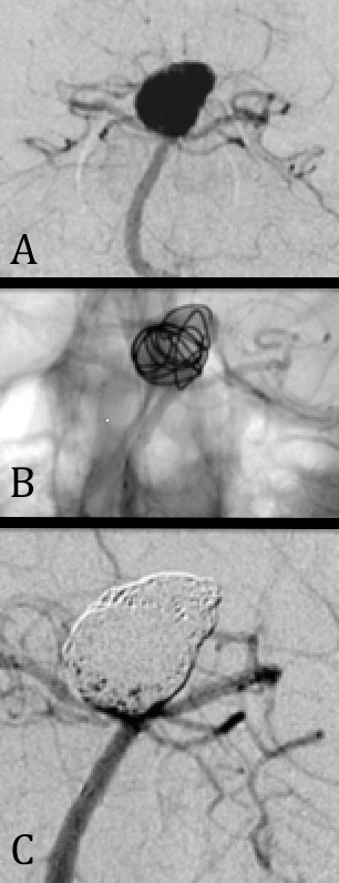
Case 2: Stent-Assisted Coiling
A: AP angiogram of an unruptured aneurysm of the basilar artery. These aneurysms are difficult to access surgically, thus endovascular coiling is often the preferred method of treatment. The aneurysm is “wide-necked” and will not safely hold coils without the assistance of a stent to maintain patency of the normal parent vessel.
B: Intra-procedural view after a specially designed stent (not visible) has been placed to protect the normal blood vessels. Endovascular coils are being deployed within the aneurysm and are held within the aneurysm by the stent. The physical nature of the coils is apparent in this image.
C: Final view of the aneurysm after it has been successfully treated with platinum coils. The aneurysm no longer fills with contrast or blood. The normal blood vessels have excellent blood flow and the stent is effectively holding the coils in place within the aneurysm.

Case 2: Stent-Assisted Coiling
Related Treatments at New Jersey Brain and Spine
At New Jersey Brain and Spine, we offer comprehensive care for cerebrovascular conditions. In addition to brain aneurysm endoscopic coiling, we provide several related treatments:
- Brain Aneurysm Clipping – A surgical alternative for aneurysm treatment
- Cerebral Angiography – A diagnostic procedure used to visualize blood vessels in the brain
- Stroke Treatment – Emergency and preventative care for stroke patients
- Vascular Malformation Treatment – Procedures for treating abnormal blood vessel formations
Frequently Asked Questions About Brain Aneurysm Endoscopic Coiling
How effective is brain aneurysm endoscopic coiling at preventing rupture?
Brain aneurysm endoscopic coiling is highly effective, with studies showing over 90% success rate at preventing initial or recurrent bleeding when performed by experienced neurosurgeons.
What are the coils made of and how do they work?
The coils are made of soft platinum and contain significant technology to ensure safety and effectiveness. They work by filling approximately 20% to 30% of the aneurysm, causing the blood inside to clot and block the aneurysm from circulation.
Will the coils remain in my brain permanently?
Yes, the platinum coils remain in the aneurysm permanently. They are biocompatible and designed to stay in place for life.
How long will I need to take off work after brain aneurysm endoscopic coiling?
Most patients can return to desk jobs within 1-2 weeks. Those with physically demanding occupations may require additional recovery time before returning to full duties.
Is brain aneurysm endoscopic coiling covered by insurance?
Most insurance plans cover brain aneurysm coiling as it is considered a medically necessary procedure for treating aneurysms. Our office staff can help verify your coverage.


