Meningioma: What a Diagnosis Really Means
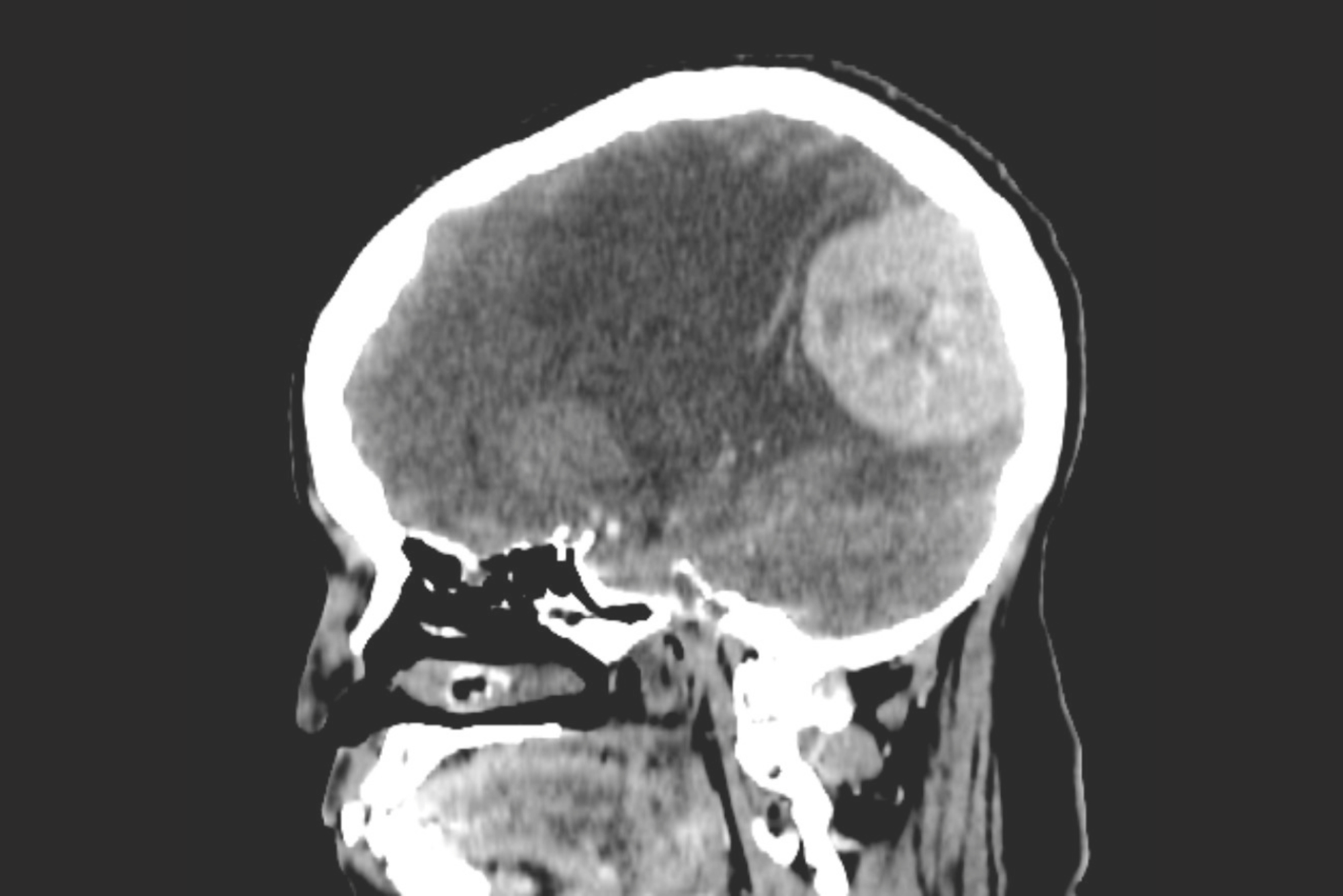
When a patient’s brain MRI shows a mass and it turns out to be a meningioma, relief and fear often arrive in the same conversation. Meningiomas are the most common primary brain tumor we see in adults, and the vast majority are benign. But “benign” is a word that needs unpacking, because what a meningioma diagnosis actually means for a given patient depends heavily on where it is, how big it is, and whether it’s causing any symptoms at all.
A meningioma doesn’t grow from brain tissue itself. It arises from the meninges, the layered membrane that wraps around and protects the brain and spinal cord. Because these tumors grow from the covering of the brain rather than from within it, they tend to push on brain tissue from the outside rather than infiltrate it, which is part of why so many meningiomas are compatible with normal, symptom-free function for years.
Most meningiomas are classified as WHO Grade I, meaning they are slow-growing and non-cancerous. A smaller number are Grade II (atypical) or Grade III (anaplastic), which tend to behave more aggressively and carry a higher risk of recurrence. Grade is determined by how the tumor cells look under a microscope after biopsy or resection, not by size or symptoms alone, which is why grade can only be confirmed with tissue, not imaging.
Location matters just as much as grade. A meningioma near the surface of the brain in a relatively “quiet” area may sit for years without causing any problem. A meningioma of the exact same size pressing on the optic nerve, a major vein, or a critical area controlling speech or movement can cause significant symptoms even while still small.
A meningioma diagnosis is not, by itself, a cancer diagnosis, and it’s not automatically a surgery diagnosis either. What actually drives our recommendation is the combination of grade, location, growth pattern over time, and whether it is producing real neurological symptoms, not the fact that a mass is visible on a scan.
Many meningiomas, particularly small ones found incidentally on imaging done for an unrelated reason like a headache workup or a minor head injury, cause no symptoms at all. When symptoms do occur, they tend to reflect the specific area of brain being compressed rather than following one universal pattern:
Because symptoms build slowly in most cases, family members often notice changes, in memory, mood, or coordination, before the patient does.
| Grade I Meningioma | Grade II (Atypical) Meningioma | Grade III (Anaplastic) Meningioma | |
|---|---|---|---|
| Definition | Slow-growing, benign tumor cells | More cellular tumor tissue with a higher recurrence risk than Grade I | Rare, malignant tumor tissue with the highest recurrence risk |
| Typical presentation | Often incidental or slowly progressive symptoms over years | Can present with more rapid symptom onset or faster growth on serial imaging | Often more rapid symptom onset and faster growth on serial imaging |
| Core causes | Sporadic cell changes in the meninges; more common with age and in women | Similar origin, but with atypical cellular features under the microscope | Similar origin, but with clearly malignant cellular features under the microscope |
| Surgical necessity | Often monitored if small and asymptomatic; surgery considered for growth or symptoms | Surgery usually recommended; radiation afterward is decided case by case based on how much tumor was removed and recurrence risk | Surgery followed by radiation is typically recommended |
For a meaningful number of patients, particularly those with small, incidentally discovered meningiomas without symptoms, our first recommendation is watchful waiting. That means a repeat MRI, typically at 6 months and then annually if stable, to confirm the tumor isn’t growing. Many meningiomas never require treatment at all.
Once a meningioma is causing symptoms, growing on serial imaging, or sitting in a location where future growth would put critical structures at risk, we shift toward active treatment. The two main options are surgical resection and stereotactic radiosurgery (such as Gamma Knife). Surgery is generally favored when a tumor is large enough to be causing symptoms and is in a location that’s surgically accessible with acceptable risk. Radiosurgery may be an option for some smaller tumors, but location matters. Tumors close to the optic nerves, for example, can limit how safely radiation can be delivered. The decision depends on tumor size, location, and radiation safety considerations.
Even after surgery, our approach depends on the grade of the tumor. A completely resected Grade I meningioma often needs nothing further beyond surveillance imaging. Radiation after surgery is more consistently recommended for Grade III tumors. For Grade II tumors, the decision depends on factors such as how much of the tumor was removed and the risk of recurrence. Incomplete removal of a Grade I tumor doesn’t automatically mean radiation is needed.
A 61-year-old woman had a small meningioma discovered incidentally after a minor car accident led to a head CT. She had no headaches, no vision changes, and a completely normal neurological exam. Her tumor measured just over a centimeter and was located along the surface of the brain away from critical structures. We recommended surveillance imaging rather than treatment, and at her one-year follow-up MRI, the tumor was unchanged.
A 54-year-old man came to us after his wife noticed several months of increasing forgetfulness and a subtle personality change, becoming more withdrawn and less engaged with family. Imaging revealed a meningioma pressing on his frontal lobe that had grown noticeably compared to a scan done a year earlier at another facility. Given the tumor’s growth and the clear cognitive symptoms it was producing, we recommended surgical resection. His memory and personality returned to baseline over the following months.
Note: These patient scenarios are representative, hypothetical examples used for illustrative purposes. Because every patient and medical history is unique, actual treatment recommendations and surgical settings depend entirely on your individual health circumstances and a thorough evaluation by your care team.
If a meningioma has already been found on imaging, a neurosurgical consultation is worthwhile even if no treatment is ultimately needed, simply to establish a monitoring plan and a clear understanding of what to watch for. You should seek evaluation, or move up an already scheduled follow-up, if you notice:
Seek emergency care immediately, rather than waiting for a follow-up visit, for:
Meningioma is common enough that we see a steady stream of patients in our New Jersey practice whose tumors were found incidentally years before they ever became symptomatic. A second opinion, especially when the recommendation is immediate surgery for a small, asymptomatic tumor, is often worth seeking, since surveillance is a legitimate and frequently appropriate path for many of these patients.
No. The large majority of meningiomas are Grade I and benign, not cancerous. A smaller subset are Grade II or III, which behave more aggressively, but even these are generally treated differently than the metastatic or primary malignant brain tumors people typically associate with the word “cancer.”
No. Small, asymptomatic meningiomas, particularly those found incidentally, are frequently monitored with periodic MRI rather than treated right away. Treatment becomes the focus once a tumor is growing, causing symptoms, or located somewhere that future growth would be risky.
It can, particularly if the tumor was in a location where complete removal wasn’t possible, or if it was a higher grade tumor. This is why we continue surveillance imaging after surgery even when the resection appears complete.
In most cases, there’s no single identifiable cause. Meningiomas are more common with age and occur more frequently in women, and prior radiation exposure to the head is a known, though less common, risk factor.
This varies significantly based on tumor size, location, and the surgical approach used. Many patients are up and walking within a day or two after surgery, though full recovery and return to normal activity is typically measured in weeks to a few months depending on the individual case.
NJBS serves patients across northern New Jersey and the greater tri-state area, with offices in Paramus, Hackensack, Montclair, Montvale, Annandale, and Englewood. No referral is required to schedule a consultation.