Limitations of MR and CT guided navigation are based on the fact that imaging is obtained prior to surgery and does not reflect subtle real time changes in the position of the brain occurring during surgery. This intra-operative distortion of the brain is termed ‘brain shift’ and varies according to the location of the tumor and other factors. Intraoperative ultrasound offers a solution to this problem by providing immediate updated information on the position and location of residual tumor (Figure 1). Ultrasound, however, is not without its limitations in that the quality of the images obtained can be obscured by artifact limiting resolution.
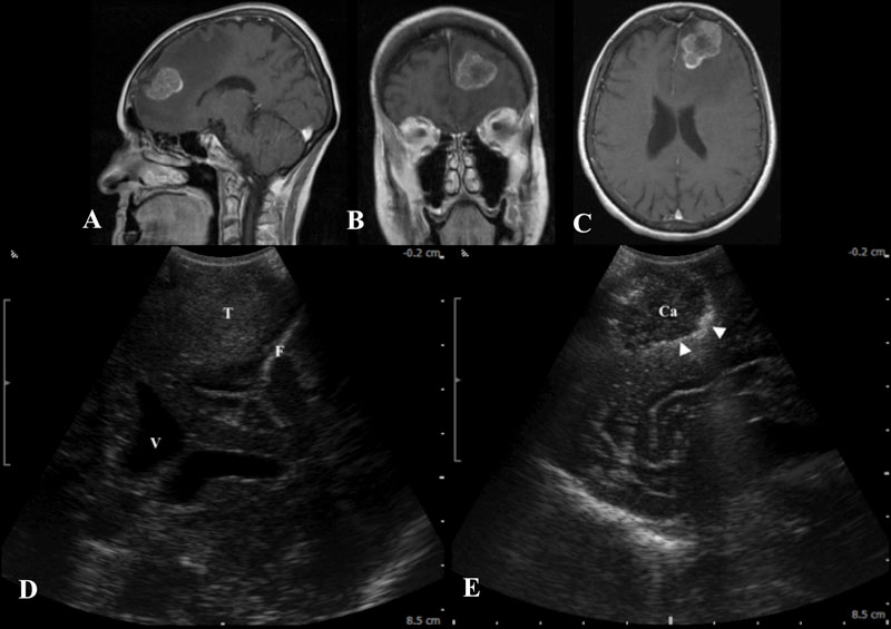
Figure 1
Figure 1: A,B,C: Pre-operative MR demonstrates a large enhancing metastasis in the left frontal lobe. These intra-operative ultrasound images show the tumor before (D) and after (E) resection in coronal and sagittal planes. Note that the post-resection image shows areas that are hyperechoic at the periphery of the tumor cavity (white arrowheads). This artifact sometimes limits the ability to determine if residual tumor exists at the periphery of the resection cavity. (T-Tumor, Ca-Tumor Cavity, V-Ventricle, F-Falx)
Have More Questions about This Condition

New Jersey Brain and Spine Earns #3 National Ranking For Neurosurgical Practices
New Jersey Brain and Spine has once again been recognized among the nation’s elite neurosurgical practices—this time...

Patient Story: Patient Gets Fitness Career Back in Shape Thanks to Dr. Roth
Robin Bray is the owner of FitnessBarre™, a popular local workout studio in Midland Park, New Jersey. The...

NJBS Highlighted for Advanced Tremor Treatment With Hackensack Meridian Health
New Jersey Brain & Spine was recently featured alongside Hackensack Meridian Health, highlighting the impact of...

Patient Story: Ashley’s Recovery After Cervical Disc Herniation
A stroke at birth left Ashley with cerebral palsy and limited mobility on her right side. So when she woke up at 1...

Back on the Competition Track: A Young Athlete’s Recovery After L4–L5 Disc Herniation Surgery
Severe nerve compression brought life to a halt—lumbar microdiscectomy helped him reclaim strength, mobility, and...

Ground-Breaking HiFu Treatment Re-Opens a Door
Innovative non-invasive essential tremor treatment puts car enthusiast back in the driver’s seat Dan lived with...
")
Full Patient Story: Chris’s Recovery After L4–5 Posterior Lumbar Interbody Fusion (PLIF)
How an L4–5 Posterior Lumbar Fusion Gave Chris Feeling in His Leg Again and His Life Back Chris was only in his early...

Back in the Game: Brian’s Recovery After C5–6 Disc Replacement
Neck pain and cervical radiculopathy stole his mobility, his golf swing, and his comfort, an artificial disc...

New Jersey Brain and Spine Launches $2000 Healthcare Scholarship to Support Next Generation of Healthcare Professionals
Investing in the future of healthcare: Annual scholarship open to high school and undergraduate students from or...