This technology has been present for over 15 years and links patient anatomy in the operating room to an imaging study obtained expressly for surgical planning immediately prior to operation. This device occasionally requires the placement of ‘fiducials‘ (Figure 1) or markers that are affixed to the patient with a gentle adhesive prior to obtaining the planning MR; these markers are identifiable on a computer monitor in surgery and allow for correlation of the patient’s head to its representation on imaging. Alternatively, the unique facial contours of a patient are defined by ‘tracing’ the face with a laser or probe thereby obviating the need to place fiducials pre-operatively.
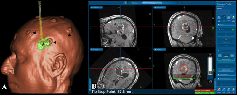
Figure 1ab
The left image ‘a’ is a rendering of a patient on a navigational workstation with fiducials indicated by arrowheads. The markers are affixed prior to the acquisition of an MR or CT and serve as localizing points visualized on imaging and allow for a correlation with real anatomy in the operating room. The asterisk indicates location of tumor. The right image ‘b’ demonstrates the proposed trajectory of a biopsy cannula in a separate patient to obtain a sample of a large deep-seated tumor.
During surgery, surgical tools are identified by optical cameras and are overlain on a monitor demonstrating the patient’s MR imaging. This tool permits more precise incisions, skull-openings (craniotomy) and dissection within the nervous system as well as guiding tumor removal. Navigational information serves as a useful adjunct but does not replace the experience and knowledge of the surgeon. (Figure 2,3).
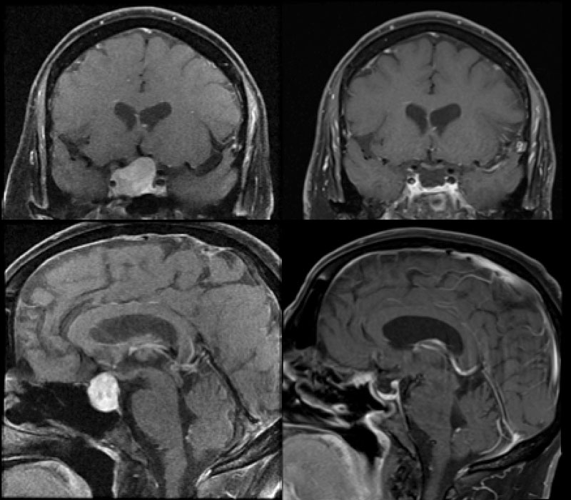
Figure 2
This 48 year old man presented with visual loss and a non-functioning pituitary tumor with lateral displacement of the medial wall of the cavernous sinus. Endonasal endoscopic surgery was performed with complete resection (left top and bottom preoperative, right top and bottom postoperative).
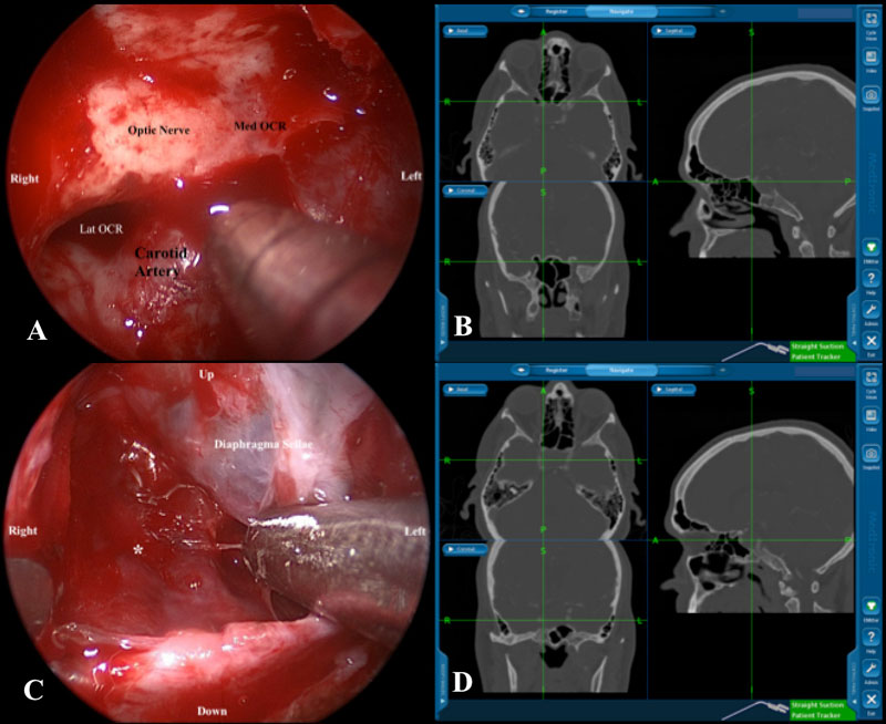
Figure 3
Intraoperative images of the case noted above demonstrate the utility of navigation. A,B: Top row with suction probe just below with optic canal and anterior to the parasellar carotid artery. C,D: Lower row with probe within the sella turcica just medial to the intra-cavernous carotid artery deep to the medial wall of the cavernous sinus (Medial OCR: Medial Optico-carotid recess, Lat OCR: Lateral Optico-carotid recess, *: Carotid artery within the cavernous sinus).
Have More Questions about This Condition

New Jersey Brain and Spine Earns #3 National Ranking For Neurosurgical Practices
New Jersey Brain and Spine has once again been recognized among the nation’s elite neurosurgical practices—this time...

Patient Story: Patient Gets Fitness Career Back in Shape Thanks to Dr. Roth
Robin Bray is the owner of FitnessBarre™, a popular local workout studio in Midland Park, New Jersey. The...

NJBS Highlighted for Advanced Tremor Treatment With Hackensack Meridian Health
New Jersey Brain & Spine was recently featured alongside Hackensack Meridian Health, highlighting the impact of...

Patient Story: Ashley’s Recovery After Cervical Disc Herniation
A stroke at birth left Ashley with cerebral palsy and limited mobility on her right side. So when she woke up at 1...

Back on the Competition Track: A Young Athlete’s Recovery After L4–L5 Disc Herniation Surgery
Severe nerve compression brought life to a halt—lumbar microdiscectomy helped him reclaim strength, mobility, and...

Ground-Breaking HiFu Treatment Re-Opens a Door
Innovative non-invasive essential tremor treatment puts car enthusiast back in the driver’s seat Dan lived with...
")
Full Patient Story: Chris’s Recovery After L4–5 Posterior Lumbar Interbody Fusion (PLIF)
How an L4–5 Posterior Lumbar Fusion Gave Chris Feeling in His Leg Again and His Life Back Chris was only in his early...

Back in the Game: Brian’s Recovery After C5–6 Disc Replacement
Neck pain and cervical radiculopathy stole his mobility, his golf swing, and his comfort, an artificial disc...

New Jersey Brain and Spine Launches $2000 Healthcare Scholarship to Support Next Generation of Healthcare Professionals
Investing in the future of healthcare: Annual scholarship open to high school and undergraduate students from or...