Degenerative Spondylolisthesis and Segmental Instability
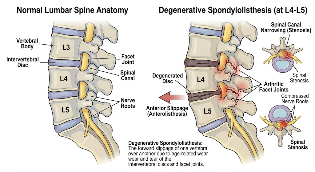
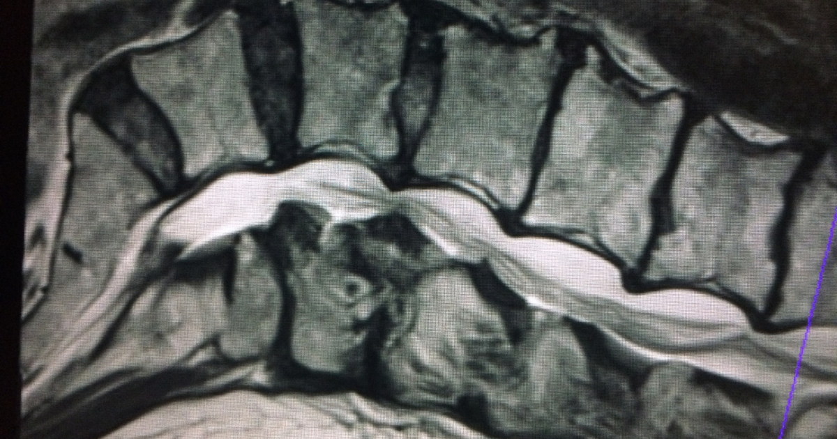
Degenerative spondylolisthesis is one of those diagnoses that tends to alarm patients more than the finding itself usually warrants. A vertebra has shifted forward, which sounds significant, but what actually determines the treatment path is whether that segment is stable and whether it’s producing symptoms that match what the patient is experiencing. Many patients are surprised to learn that the amount of slippage is often less important than whether that slippage is stable or still moving.
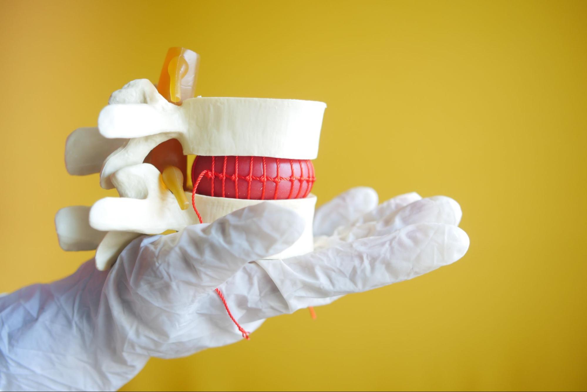
Unlike isthmic spondylolisthesis, which stems from a fracture in the bony pars interarticularis, degenerative spondylolisthesis develops from years of wear on the facet joints and disc.
As the disc loses height and the facet joints (the small stabilizing joints at the back of each spinal segment) develop arthritic changes, the normal restraints that keep one vertebra aligned over the next gradually loosen. Over time, this allows the upper vertebra to slide forward relative to the one below it, most commonly at L4-5. This pattern typically develops later in life and is often accompanied by spinal stenosis, since the same degenerative process that allows the slippage also narrows the canal.
The presence of a slip alone doesn’t answer the most important clinical question, which is whether that segment is unstable.
A 10% slip that hasn’t changed in five years is a fundamentally different clinical problem than a 10% slip that’s actively worsening between flexion and extension.
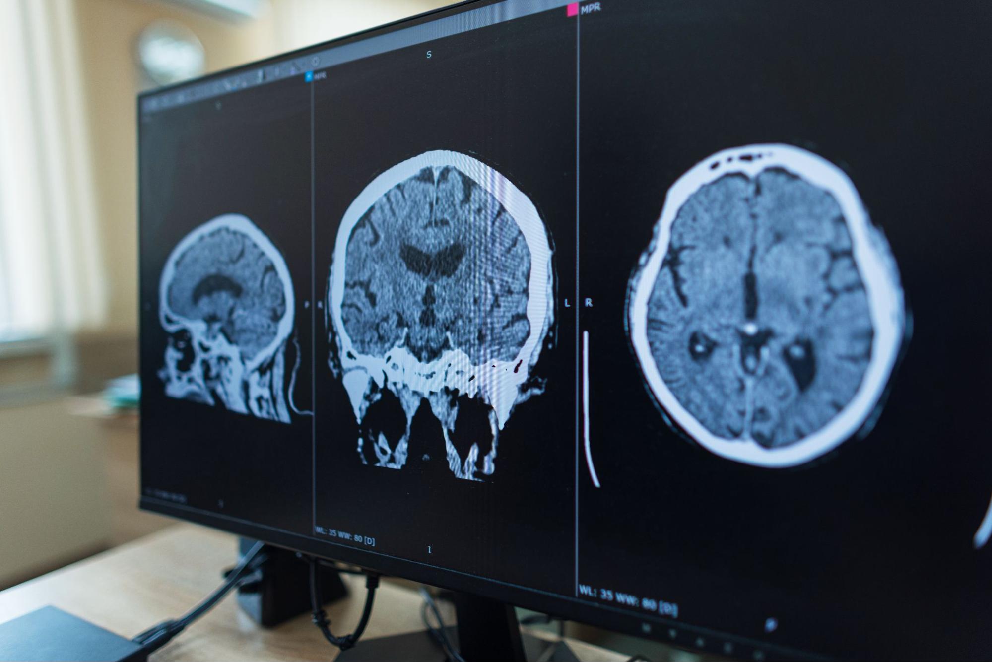
We assess instability using flexion-extension X-rays, which allow us to compare how the vertebra moves when you bend forward and backward. Unlike an MRI, which captures the spine in a single position, these X-rays help us determine whether the vertebra is actively moving between positions. Meaningful movement indicates dynamic instability, meaning the segment is still moving in a way it shouldn’t. A slip that remains unchanged regardless of position is generally considered more stable, even at a similar grade.
Dynamic instability is more likely to contribute to significant mechanical back pain, while nerve-related symptoms such as radiculopathy or neurogenic claudication can occur with either stable or unstable spondylolisthesis when associated stenosis is present.
Key mechanical factors we weigh:
| Feature | Stable Slip | Unstable (Dynamic) Slip |
|---|---|---|
| Flexion-extension X-ray findings | Minimal change in slip percentage between positions | Significant change in slip percentage between positions |
| Typical symptoms | Mechanical low back pain, may be mild | Often combined with neurogenic claudication from associated stenosis |
| Progression over time | Often remains unchanged for years | More likely to progress if untreated |
| Surgical necessity | Usually managed conservatively unless symptoms are significant | More often requires decompression with fusion if conservative care fails |
At NJBS, we don’t recommend surgery based solely on the amount of slippage. We consider whether the segment is stable, how much it affects your daily function, whether nerves are being compressed, and how you’ve responded to conservative treatment. Those factors together help determine whether continued observation, decompression, or decompression with fusion is the most appropriate option.
A 66-year-old woman had a Grade 1 degenerative spondylolisthesis at L4-5 discovered on imaging for mild, intermittent low back pain. Flexion-extension X-rays showed no meaningful instability, and she had no leg symptoms. She was managed with a core-strengthening physical therapy program and activity modification, with good symptom control over time.
A 71-year-old man presented with progressive walking limitation from neurogenic claudication, needing to stop every block or two. Imaging showed a Grade 2 degenerative spondylolisthesis at L4-5 with associated stenosis, and flexion-extension films confirmed clear segmental instability. He had already completed physical therapy and two epidural injections with only temporary benefit. Given the confirmed instability, stenosis, and exhausted conservative options, he underwent decompression with fusion at L4-5, and his walking tolerance improved substantially afterward.
Note: These patient scenarios are representative, hypothetical examples used for illustrative purposes. Because every spine and medical history is unique, actual treatment recommendations and surgical settings depend entirely on your individual health circumstances and a thorough evaluation by your care team.
Evaluation is worth pursuing when:
Cauda equina syndrome is a surgical emergency. Saddle numbness, loss of bowel or bladder control, or progressive weakness in both legs requires immediate emergency room evaluation or a call to 911.
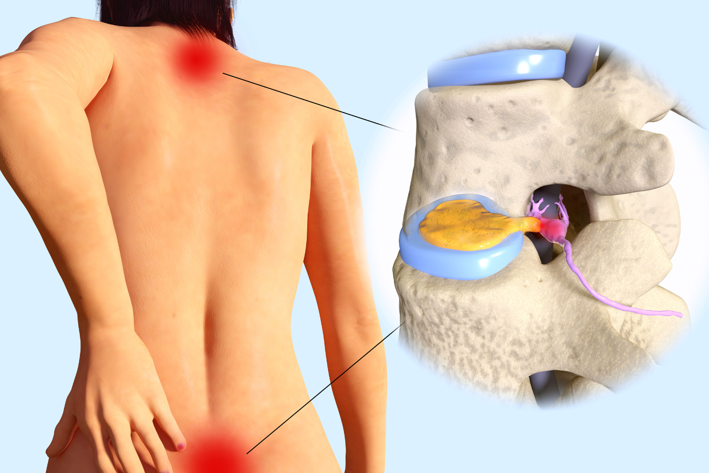
No. It’s a slippage of one vertebra over another from facet and disc wear, whereas a herniated disc involves the annulus tearing and disc material displacing. They can occur together but are distinct problems.
Not necessarily. Many slips remain stable for years, which is part of why we recheck flexion-extension imaging rather than assuming automatic progression.
Not automatically. Many patients with mild instability and manageable symptoms do well with conservative care. Fusion becomes a more serious consideration when instability is confirmed alongside significant, persistent functional limitation.
The same degenerative process, facet and disc wear, that allows the vertebra to slip forward often also narrows the spinal canal, which is why the two conditions frequently occur together.
NJBS serves patients across northern New Jersey and the greater tri-state area, with offices in Paramus, Hackensack, Montclair, Montvale, Annandale, and Englewood. No referral is required to schedule a consultation.