Why Do I Have New SI Joint Pain After Lumbar Fusion?
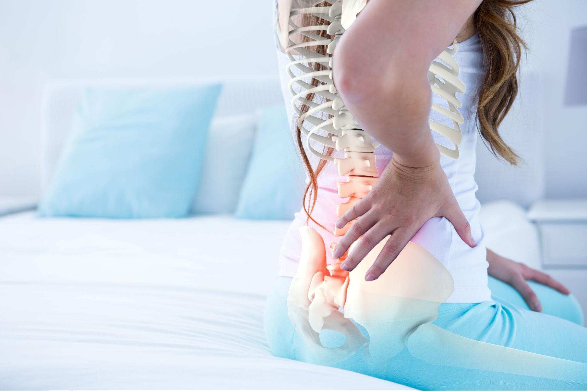
A common pattern shows up in follow-up visits after a lumbar fusion: a patient’s original back and leg pain is gone, but a new, different pain has developed low in the back or buttock that wasn’t there before. It’s a distinct complaint, often felt more to one side, and it tends to worsen with sitting, standing from a seated position, or rolling over in bed. In many of these patients, the pain generator isn’t the fusion itself. It’s the sacroiliac (SI) joint just below it.
When new pain develops after lumbar fusion, the first step is determining where it’s actually coming from. Sometimes the source is the spine itself. Other times, it may be the adjacent levels or another structure entirely, such as the sacroiliac (SI) joint.
The SI joints sit where the base of the spine (the sacrum) meets the pelvis (the ilium) on each side. Unlike the disc spaces we fuse in the lumbar spine, the SI joints are true joints, with small amounts of motion, held together by some of the strongest ligaments in the body. In a normally functioning spine, motion and load are distributed across many segments, including these joints.
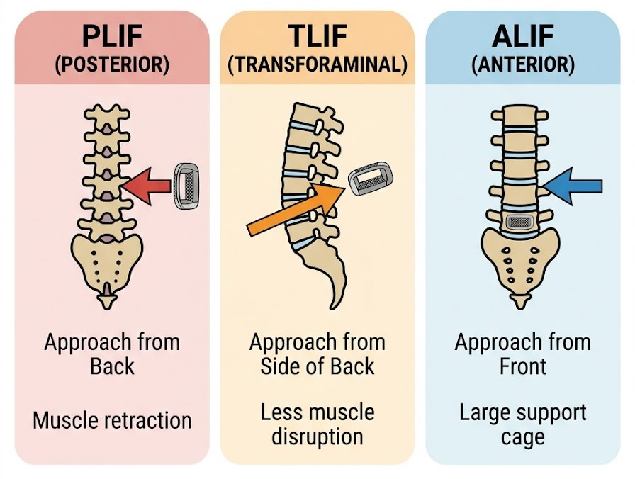
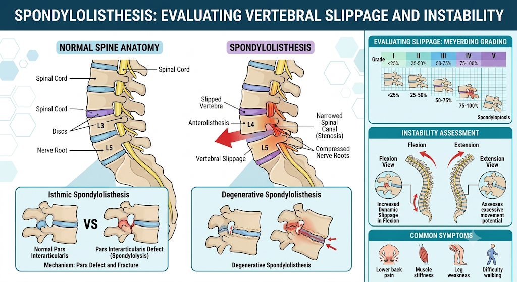
When a segment like L4-S1 is instrumented and fused, that segment stops moving by design. That’s the point of the surgery. But the body still needs to bend, twist, and transfer load somewhere. The segments directly above a fusion tend to take on extra motion, which is why we talk about adjacent segment changes at those levels. Less commonly discussed, but very real clinically, is that the SI joints below a lumbosacral fusion can absorb a similar increase in shear and rotational force, especially when the fusion extends all the way to the sacrum or includes pelvic fixation with iliac screws.
When we fuse the lumbosacral junction, we’re not eliminating motion from the system. We’re relocating it. The SI joint is often the next available segment to take on that load.
A few mechanical points worth understanding:
Because both conditions can cause low back and buttock pain after a fusion, telling them apart matters for treatment planning.
| Feature | SI Joint Pain (Post-Fusion) | Recurrent Lumbar Pathology (Adjacent Segment) |
|---|---|---|
| Typical location | Low back/buttock, usually one-sided, below the fusion | Low back, may radiate into the leg, at or above the fusion |
| Provoking movements | Transitional movements (sit to stand), prolonged sitting, stairs | Extension-based movements, prolonged standing/walking |
| Exam findings | Positive FABER test, Gaenslen’s test, tenderness over the joint itself | Positive extension-based provocative signs, possible neurological findings |
| Imaging | Fusion construct intact; SI joint may show degenerative change | New stenosis, disc degeneration, or instability at the level above the fusion |
| Diagnostic confirmation | Image-guided SI joint injection with significant relief | Correlating imaging with the exam; sometimes a diagnostic nerve block |
When a patient comes to us after a fusion done elsewhere (or one we performed ourselves) with new pain that doesn’t match their original symptoms, we don’t assume it’s a failed fusion. We re-examine the whole kinetic chain, including the SI joints, and we let the exam and confirmatory imaging guide us rather than assuming the new pain must originate from the hardware itself. This is a scenario we see regularly among New Jersey patients who’ve had a fusion performed elsewhere and are seeking a second opinion on new symptoms.
A patient in her late 50s had an L4-S1 instrumented fusion for degenerative disc disease. Her leg pain resolved well, but about eight months later she developed new, one-sided low back pain that was worse getting up from a chair. Flexion-extension X-rays showed a solid, well-positioned fusion with no hardware concerns. On exam, she had a positive FABER test and tenderness directly over the SI joint. An image-guided diagnostic injection into the joint gave her near-complete, though temporary, relief, confirming the joint as the source. She was started on a targeted physical therapy program focused on SI joint stabilization and pelvic mechanics, along with a therapeutic injection series. Her symptoms improved significantly over the following months, and she did not require further surgery.
A patient in his early 60s had undergone a longer L3-S1 fusion with iliac screw fixation for adult degenerative scoliosis. Roughly a year out, he developed persistent, disabling SI joint pain confirmed on two separate diagnostic blocks. He completed an extensive course of physical therapy and a series of therapeutic injections without lasting relief. Given the confirmed pain generator and the failure of conservative measures, he was ultimately a candidate for minimally invasive SI joint fusion, which he elected to pursue, with meaningful improvement in his symptoms afterward.
Note: These patient scenarios are representative, hypothetical examples used for illustrative purposes. Because every spine and medical history is unique, actual treatment recommendations and surgical settings depend entirely on your individual health circumstances and a thorough evaluation by your care team.
New or different pain after a fusion is worth having evaluated, particularly if it:
Cauda equina syndrome is a surgical emergency. If you experience saddle numbness, loss of bowel or bladder control, or progressive weakness in both legs at any point after a fusion, this requires immediate emergency room evaluation or a call to 911. Do not wait for a scheduled office visit.
Not necessarily. A fusion can be solid and well-positioned on imaging while the SI joint develops pain due to the change in how load moves through the pelvis. These are two separate issues, and we evaluate them separately.
The most reliable way is an image-guided diagnostic injection directly into the joint. If that injection significantly relieves the pain, at least temporarily, it strongly supports the SI joint as the generator.
It’s a recognized phenomenon, particularly after fusions that extend to the sacrum or include pelvic fixation, though not every patient develops it. Individual anatomy and pre-existing wear play a role.
Physical therapy focused on pelvic and SI joint mechanics, activity modification, and a course of image-guided injections are standard first steps. Surgery is only considered when these measures fail to provide lasting relief.
NJBS serves patients across northern New Jersey and the greater tri-state area, with offices in Paramus, Hackensack, Montclair, Montvale, Annandale, and Englewood. No referral is required to schedule a consultation.