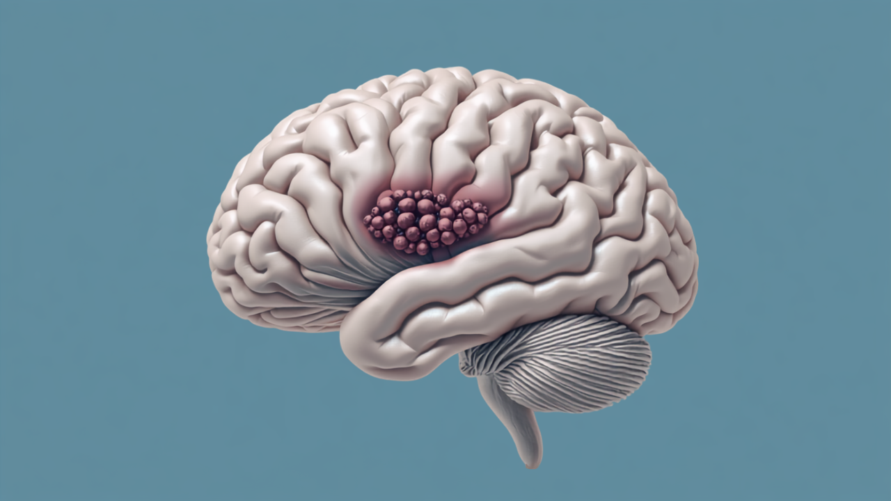
Brain Tumors: How Location Determines Symptoms and Treatment
Where a brain tumor is located tells us nearly as much as what type of tumor it is. The symptoms a patient experiences, the degree of functional risk surgery carries, the structures that must be protected during any intervention, and the overall prognosis are all shaped substantially by the tumor’s position within the brain. Two patients with the same tumor histology and the same grade can present in entirely different ways, follow entirely different treatment courses, and require entirely different surgical approaches because their tumors occupy different anatomical territories.
At New Jersey Brain and Spine, tumor location is one of the first things we consider when a patient comes to us with a new brain tumor diagnosis. It is not the only consideration, but it is foundational. Understanding what each region of the brain controls, and what happens when a tumor disrupts that region, allows both our clinical team and our patients to approach diagnosis and treatment planning with greater clarity.
The brain is not a homogeneous structure. Different regions are responsible for different neurological functions, and the relationship between anatomy and function is reasonably predictable. This means that the symptoms produced by a brain tumor tend to reflect the role of the region being affected, whether through direct tissue invasion, compression of adjacent structures, edema (swelling), or disruption of local neural circuitry.
Some symptoms are region-specific: a tumor in the occipital lobe may cause visual field defects because the occipital lobe processes visual information, while a tumor in the motor cortex of the frontal lobe may produce weakness on the opposite side of the body. Other symptoms are less specific and reflect increased intracranial pressure rather than local effects: persistent headache (often worse in the morning), nausea, vomiting, and cognitive slowing can occur with tumors in multiple locations when they produce significant mass effect or obstruct cerebrospinal fluid circulation.
The major functional regions of the brain most relevant to tumor-related symptoms are the frontal lobe (executive function, personality, motor control, expressive speech), the temporal lobe (memory, language comprehension, hearing), the parietal lobe (sensory processing, spatial awareness, reading and calculation), the occipital lobe (visual processing), the cerebellum (coordination, balance, fine motor control), and the brainstem (breathing, heart rate, swallowing, eye movements, arousal).
| Brain Region | Key Functions | Symptoms a Tumor in This Region May Cause |
|---|---|---|
| Frontal lobe | Planning, personality, motor control, expressive speech (Broca’s area) | Personality or behavior changes, impaired judgment, weakness on one side of the body, difficulty producing speech |
| Temporal lobe | Memory formation, language comprehension, hearing | Memory loss, difficulty understanding spoken or written language, hearing changes, seizures with unusual auras |
| Parietal lobe | Sensory processing, spatial awareness, reading, calculation | Numbness or tingling on one side, difficulty reading or doing math, spatial disorientation, neglect syndrome |
| Occipital lobe | Visual processing | Visual field loss, visual disturbances, difficulty recognizing objects or faces |
| Cerebellum | Balance, coordination, fine motor control | Ataxia (unsteady gait), loss of coordination, tremor, slurred speech, difficulty with fine movements |
| Brainstem | Breathing, heart rate, swallowing, eye movements, arousal | Double vision, facial weakness or numbness, difficulty swallowing, hearing changes, limb weakness, coordination problems |
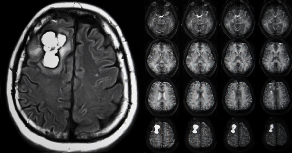
This patient was referred after an MRI obtained for unrelated headaches revealed a 2.1 cm extra-axial mass with the imaging characteristics of a meningioma arising from the frontal convexity. She was neurologically intact. Her personality and executive function were unaffected. She had no seizures and no motor deficits.
Meningiomas are generally slow-growing tumors arising from the meninges rather than the brain itself. Given her intact neurological status, the benign-appearing imaging characteristics, and the tumor’s location in a region with limited immediate functional risk at its current size, her care team recommended surveillance with serial MRI imaging rather than immediate surgery. She was counseled about the symptoms that would prompt reassessment: new headaches, personality changes, weakness, or seizure activity.
At 18-month follow-up, the tumor remained stable in size with no new symptoms. Observation continues.
This patient presented with new-onset seizures and a two-week history of progressive word-finding difficulty. MRI revealed a heterogeneously enhancing mass in the left temporal lobe with surrounding edema and mass effect on adjacent structures. The imaging features were highly concerning for high-grade glioma.
Given the tumor’s location near Wernicke’s area (the region responsible for language comprehension) and the progressive neurological deficit, surgical planning required careful consideration of language preservation. Functional MRI and intraoperative neurophysiological monitoring were used to map language function and guide the extent of resection. Maximal safe resection was performed, followed by adjuvant radiation and chemotherapy. His word-finding deficits partially recovered with speech therapy in the postoperative period.
Note: These patient scenarios are representative, hypothetical examples used for illustrative purposes. Because every case involves unique tumor biology, location, and patient circumstances, actual treatment recommendations depend entirely on a thorough individual evaluation by a qualified neurosurgical care team.
A new diagnosis of brain tumor should be evaluated by a neurosurgeon with specific experience in neuro-oncology. The neurosurgical assessment addresses tumor characteristics (size, location, imaging appearance, rate of growth), functional risk (proximity to eloquent cortex, white matter tracts, or critical vessels), and the appropriate role of surgery within a multimodal treatment plan that may also include radiation therapy, chemotherapy, or immunotherapy.
Patients in New Jersey who have received a brain tumor diagnosis or who are experiencing symptoms that may warrant imaging evaluation are encouraged to seek consultation at NJBS. Early evaluation allows the full range of diagnostic and treatment options to be considered at a point when they are most likely to be beneficial.
Some brain tumor presentations require emergency evaluation and should not wait for a scheduled appointment. Call 911 or go to the nearest emergency room immediately if you experience sudden onset of severe headache that is the worst of your life (particularly if accompanied by neck stiffness or photophobia), sudden loss of consciousness, acute onset of arm or leg weakness or numbness on one side of the body, sudden inability to speak or understand speech, new onset seizure, or sudden vision loss.
These presentations can indicate acute hemorrhage into a tumor, herniation due to rapidly increasing intracranial pressure, or other neurological emergencies requiring immediate intervention.
Not necessarily. A small tumor in a functionally critical area, such as the brainstem, the motor cortex, or Broca’s or Wernicke’s language areas, can produce dramatic symptoms. A larger tumor in a relatively silent region, such as the right frontal pole in a right-handed individual, may produce few or no symptoms for a long period. Symptom severity is determined by the tumor’s functional impact: how much pressure it is generating, whether it is infiltrating critical structures, and whether it is producing edema. Size matters, but location matters more for the clinical picture.
Yes, particularly when the tumor involves the frontal lobe or its connections. The frontal lobes are responsible for executive function, impulse control, social judgment, and personality regulation. Patients with frontal lobe tumors or tumors that affect frontal lobe connectivity may become more impulsive, lose insight, display irritability or emotional blunting, or show changes in motivation and social behavior. Family members often notice these changes before the patient does, which is why a detailed history from someone who knows the patient well is a valuable part of the clinical evaluation.
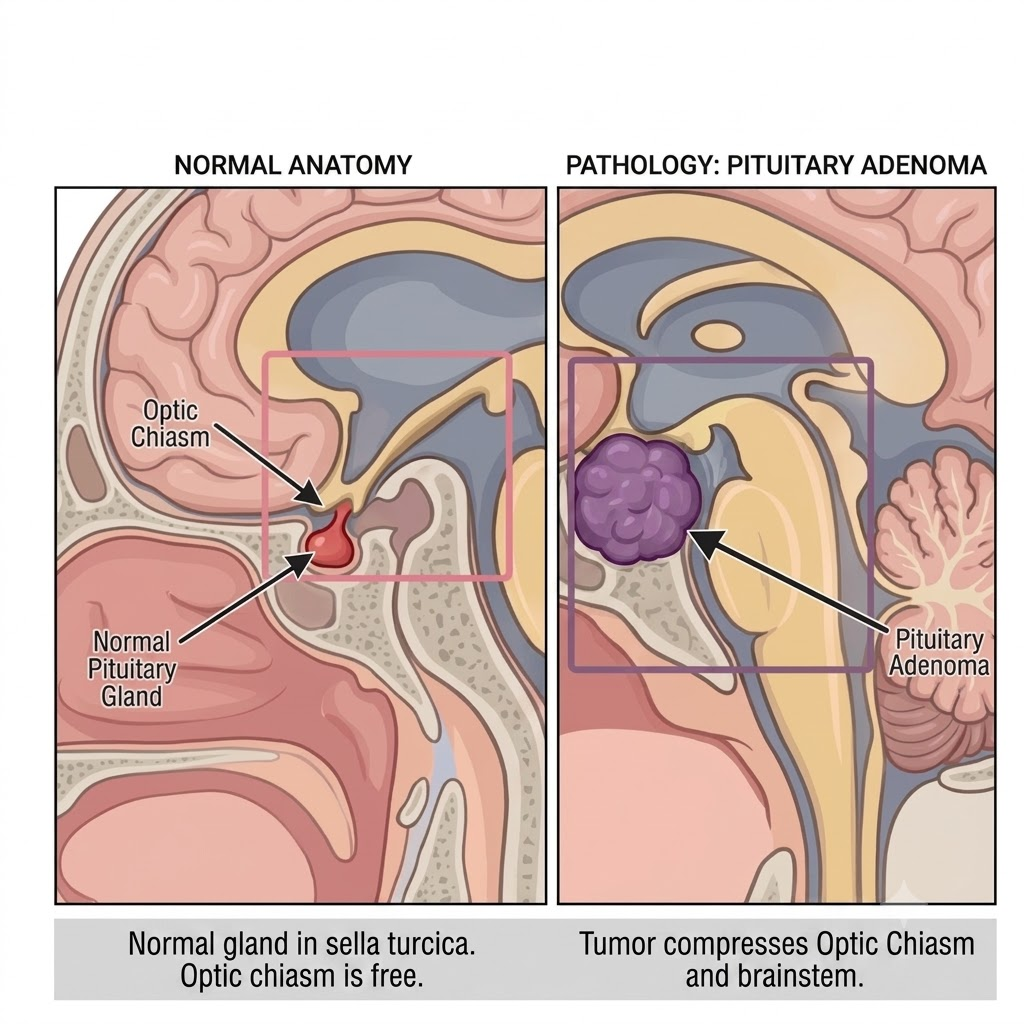
A primary brain tumor originates in the brain tissue itself or in the structures lining the brain (such as the meninges). Examples include gliomas, meningiomas, schwannomas, and pituitary adenomas. A metastatic brain tumor originates elsewhere in the body and spreads to the brain through the bloodstream. Common sources include lung, breast, melanoma, colon, and kidney cancers. The two types have different biological behaviors, different treatment strategies, and different prognoses, though both can produce symptoms based on their location and mass effect.
For tumors near eloquent cortex (regions responsible for language, motor function, or sensory processing), neurosurgeons may use intraoperative mapping techniques to identify the exact location of critical functions at the time of surgery. This can involve awake craniotomy, in which the patient is kept conscious during portions of the procedure to allow real-time language or motor testing, along with electrical stimulation mapping of the cortical surface. The goal is to achieve the maximum extent of tumor resection while preserving the functional tissue that determines the patient’s quality of life. At NJBS, intraoperative neurophysiological monitoring is integrated into surgical planning for tumors in functionally critical locations.
Brain tumors are graded by the World Health Organization (WHO) on a scale from 1 to 4 based on histological and molecular features that predict biological behavior. Grade 1 and 2 tumors are generally slow-growing, while Grade 3 and 4 tumors are more aggressive. Glioblastoma (GBM), the most common malignant primary brain tumor in adults, is WHO Grade 4 and carries a significantly more guarded prognosis than lower-grade gliomas. However, grading is just one element of prognosis: molecular markers, patient age and functional status, extent of resection, and response to adjuvant therapy all contribute to outcomes. Treatment decisions at NJBS incorporate the full molecular and clinical profile, not grade alone.
NJBS serves patients across northern New Jersey and the greater tri-state area, with offices in Paramus, Hackensack, Montclair, Montvale, Annandale, and Englewood. No referral is required to schedule a consultation.