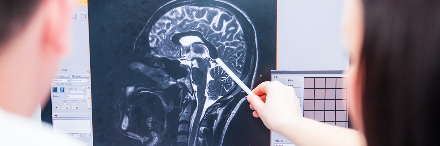
Conditions
For over 20 years, the experienced physicians at New Jersey Brain and Spine have successfully treated the full array of neurosurgical conditions, and more than 40,000 patients have benefited from our care. Whether you have a complex and rare condition or a common problem like back or spine pain, our specialists can provide you with the most advanced, least invasive care to help you get better, faster.
Discover Conditions
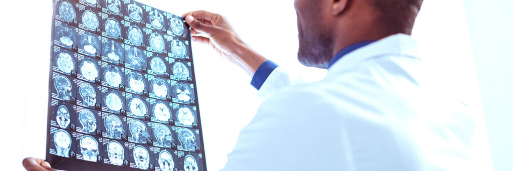
Treatments
Our physicians and surgeons are skilled in performing highly complex, proven treatments for a wide range of neurosurgical conditions. Each specialist combines advanced training with years of experience in their subspecialty area to give you exceptional results. While we perform more complex neurosurgical procedures than any other area practice, we start with the most conservative treatment option appropriate for your condition and help you fully understand all of your treatment options.
Discover Treatments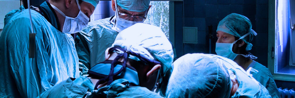
Clinical Trials
Keep up-to-date with the most recent clinical trials in which we are participating.
Discover Clinical Trials