The Truth About Pinched Nerves: Symptoms People Ignore Until Weakness or Numbness Becomes Permanent
It is a common scenario: you wake up with an acute stiffness in your neck or a deep, localized ache in your lumbar spine. It is easy to attribute the discomfort to a poor sleeping posture or natural aging, consume an over-the-counter analgesic, and continue with your daily routine.
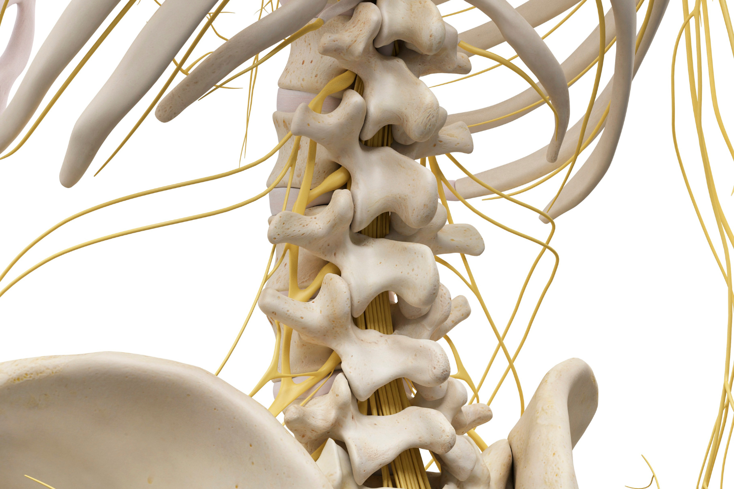
When a nerve root exiting the spinal column becomes structurally compressed or irritated — a clinical condition known as radiculopathy — pain is typically the initial symptom to manifest. Because this pain can fluctuate based on positioning, many individuals delay seeking medical care.
However, the clinical truth is unforgiving: while the musculoskeletal system can tolerate significant temporary strain, nerve roots are highly sensitive, delicate structures. Ignoring the progressive warning signs of a pinched nerve from a disc or bone spur can allow a manageable condition to quietly transition into irreversible neurological deficits.
At New Jersey Brain and Spine (NJBS), our board-certified neurosurgeons evaluate nerve compression cases daily. Identifying how a pinched nerve progresses, recognizing the subtle sensory changes that demand attention, and initiating timely care are critical components of protecting your long-term mobility.
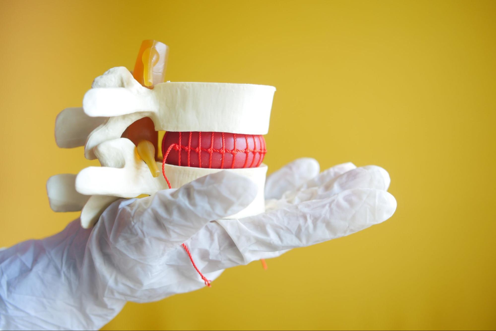
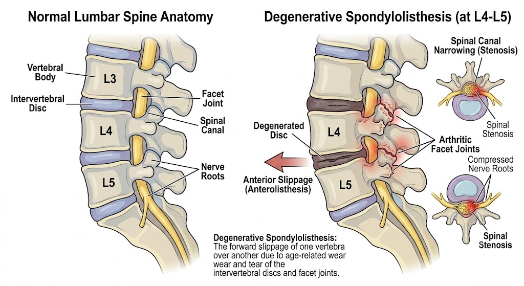
To understand the long-term risks of a pinched nerve, it helps to look at how the spine works behind the scenes. Think of the spinal cord as your body’s main electrical highway. It sends out smaller, branching nerve roots through tiny bony tunnels in the spine, which control everything from the strength in your muscles to the sensations in your arms and legs.
A pinched nerve happens when nearby tissue presses against or squeezes one of these delicate nerve paths. This pressure is usually caused by common spinal conditions, such as:
When a nerve root is subjected to constant, ongoing pressure, that physical force cuts off the flow of the tiny blood vessels that feed and nourish the nerve fibers. Starved of vital oxygen and nutrients, the nerve quickly becomes swollen, irritated, and highly inflamed. This internal distress causes the nerve to misfire, sending urgent signals of pain, burning, or tingling straight to your brain.
While symptoms vary from patient to patient, many cases of progressive nerve compression follow a pattern that begins with pain and may eventually lead to numbness or weakness if the underlying compression is not addressed. Identifying where your symptoms fall on this pathological timeline is critical for preserving long-term nerve function.
This is the symptomatic window that most patients focus on. It presents as a burning sensation down the arm, a deep ache near the scapula, or the classic shooting leg pain associated with lumbar radiculopathy (sciatica). Because this pain frequently shifts or abates with postural changes, patients often assume the underlying lesion is resolving. In reality, the mechanical pressure remains, and the nerve is actively signaling that it is in a state of acute irritation.
When acute pain begins to dull and is replaced by a constant “pins and needles” sensation (paresthesia) or a cold, diminished sensation (hypoesthesia), patients frequently misinterpret this change as a sign of clinical improvement.
From a neurosurgical standpoint, this is a dangerous misconception. The reduction in pain combined with expanding numbness indicates that the nerve fibers are losing their ability to transmit sensory data altogether. It is the anatomical equivalent of a communication line losing its signal.
This represents the clinical tipping point. When mechanical compression becomes severe enough to block the motor axons, the electrical signals can no longer reach the target muscle groups. Patients begin experiencing objective motor weakness, manifesting as dropping objects, difficulty turning door handles, an unstable knee, or a structural inability to clear the foot while walking.
While mild, intermittent aches can often be managed with conservative therapies, certain “red flag” symptoms indicate that a nerve is in critical distress and needs attention right away. You should seek an immediate specialist evaluation if you notice progressive muscle weakness, such as unexpectedly dropping objects, losing your grip, or experiencing foot drop (where your foot drags or catches as you walk). Furthermore, a sudden loss of balance, rapidly spreading numbness, or any new loss of bowel or bladder control are clear medical indicators that require urgent intervention to protect your long-term mobility.
While peripheral nerve roots possess a capacity for axonal regeneration, they have a biological point of no return. If a nerve root is subjected to high-grade, unremitting compression for a prolonged period — typically several months — the internal nerve strands (axons) undergo a process known as Wallerian degeneration.
Once these structural pathways degrade, the muscle tissues they innervate begin to waste away from a lack of neurological stimulation, a process known as neurogenic atrophy.
Even if an operative decompression is executed later to remove the underlying bone spur or herniated disc material, a severely atrophied nerve may fail to regenerate fully. This can leave the patient with permanent sensory loss or a lifelong functional deficit, such as a permanent foot drop. Waiting for physical weakness to manifest before seeking a specialist consultation is a significant risk to your long-term mobility.
A 47-year-old male presented with neck pain radiating into his right shoulder and arm after several months of worsening symptoms. Initially, he experienced intermittent pain and tingling in his thumb and index finger, which he attributed to sleeping in an awkward position. Over time, the tingling became constant and was eventually accompanied by weakness when lifting objects with his right arm.
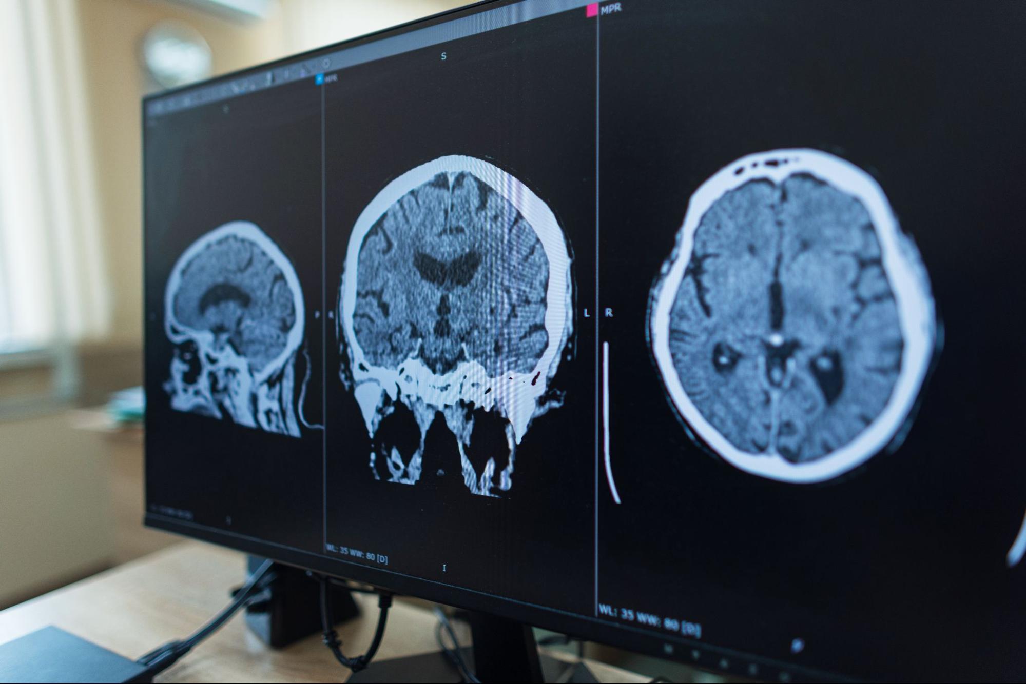
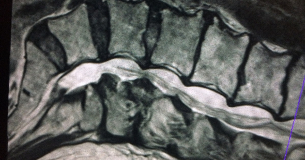
After a comprehensive neurological examination and MRI review, NJBS physicians identified a cervical disc herniation causing compression of the C6 nerve root. Because symptoms had progressed despite physical therapy, anti-inflammatory medications, and activity modification, continued observation was no longer recommended.
The patient underwent a targeted surgical decompression to relieve pressure on the affected nerve. Following treatment, his arm pain resolved, strength gradually returned, and he was able to resume normal daily activities. This case highlights the importance of early evaluation when symptoms progress from pain alone to persistent numbness or weakness.
The advantage of identifying a compressed nerve early is that it grants access to highly effective, non-operative treatment modalities. The primary objective of the NJBS neurosurgeons is to decompress the nerve root before structural, permanent nerve damage takes place.
Our clinical team utilizes an integrated, evidence-based approach to resolve radiculopathy symptoms:
There is no universal timeline because axonal degradation depends entirely on the severity and velocity of the mechanical compression. While mild, intermittent pressure can be tolerated for several months, severe and absolute compression can cause irreversible nerve fiber death within a matter of weeks. As a clinical standard, any constant numbness or objective motor weakness lasting longer than 4 to 6 weeks requires an immediate, formal evaluation by a specialist.
This transition is typically an indicator of advancing neurological decline rather than clinical healing. When a nerve root is irritated but functionally intact, it transmits rapid pain signals. If the mechanical compression escalates to a point where it compromises the nerve’s internal blood supply, the nerve loses its capacity to conduct electrical signals entirely. This causes the pain to fade, while numbness takes its place.
Yes. Foot drop is a specific motor weakness characterized by an inability or significant difficulty dorsiflexing the foot (lifting the front part of the foot toward the shin). This deficit causes patients to drag their toes or lift their knee abnormally high while walking to avoid tripping. It is a classic sign of severe compression of the L4 or L5 nerve roots in the lumbar spine and represents a critical medical milestone that requires urgent neurosurgical intervention.
If radiculopathy symptoms are mild and intermittent, specialized physical therapy can be highly effective by optimizing spinal alignment, reducing adjacent muscle spasms, and introducing nerve-gliding exercises to relieve local tension. However, if the sensory numbness is constant or accompanied by motor weakness, physical therapy alone is rarely sufficient to override a fixed mechanical blockage, such as an extruded disc fragment or a calcified bone spur.
The differentiation is defined by dermatomal anatomy. A pinched nerve originating in the cervical spine (neck) will send radiating symptoms downward through the shoulder, scapula, arm, hand, or specific fingers. Conversely, a pinched nerve rooted in the lumbar spine (lower back) will project symptoms through the gluteal region, hip, anterior or posterior thigh, calf, and down into the ankle or foot.
NJBS serves patients across northern New Jersey and the greater tri-state area, with offices in Paramus, Hackensack, Montclair, Montvale, Annandale, and Englewood. No referral is required to schedule a consultation.