Decompression vs. Spinal Fusion: How Surgeons Evaluate Stability

When a patient presents with severe back or leg pain that has proven refractory to comprehensive conservative management, surgical intervention may be indicated. When reviewing advanced neuroimaging, patients frequently encounter two core surgical categories: a standalone decompression (such as a microdiscectomy or laminectomy) and a spinal fusion.
Both procedures are highly effective at relieving nerve root compression, but they serve entirely different structural purposes. The fundamental factor that dictates which operation a specialist chooses is spinal stability.
At New Jersey Brain and Spine (NJBS), our board-certified neurosurgeons evaluate the spine not just as a collection of static images, but as a dynamic, load-bearing system. Understanding how our team objectively measures spinal stability helps clarify why a standalone spinal decompression surgery is ideal for some patients, while others require the structural reinforcement of an instrumented fusion.
To understand how neurosurgeons choose between these procedures, it helps to establish what each operation physically alters within the anatomy of the spine.
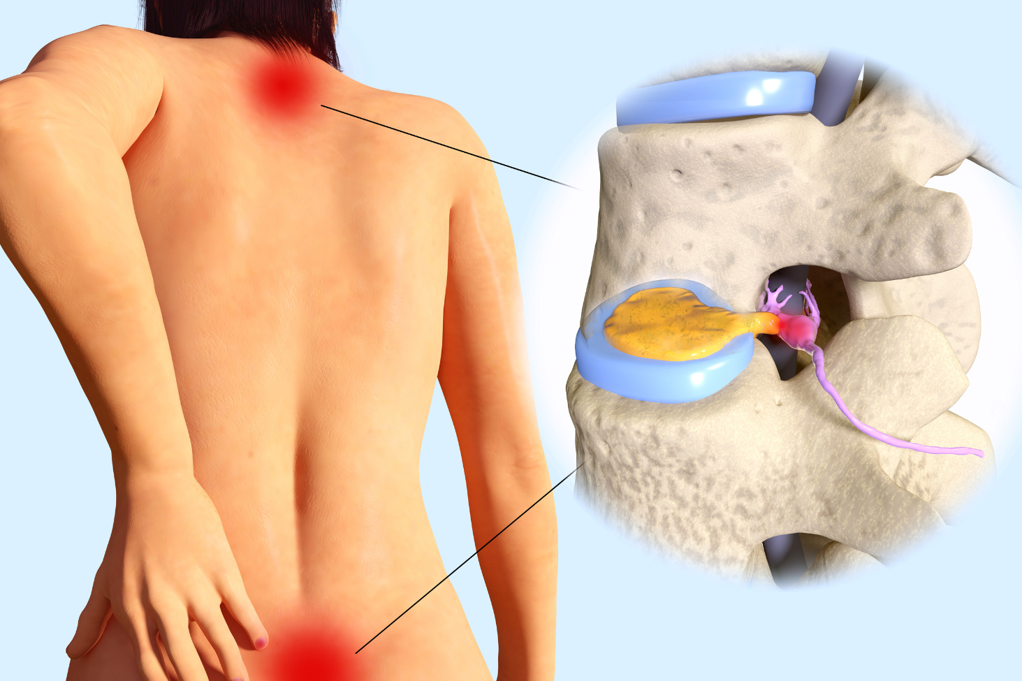
The primary objective of a decompression surgery — such as a laminectomy, microdiscectomy, or foraminotomy — is to remove a localized tissue mass that is impinging upon the spinal cord or exiting nerve roots.
During this procedure, an NJBS neurosurgeon carefully excises a specific fragment of a herniated disc, a thickened ligament, or an arthritic bone spur. A standalone decompression is a subtractive procedure; it frees the pinched nerve root by removing a minor portion of local tissue, without altering the overall structural architecture of the bony vertebral column.
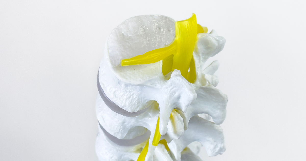
A spinal fusion, or arthrodesis, is an additive and stabilizing procedure. While it still involves decompressing pinched nerves, its primary structural goal is to permanently lock two or more vertebrae together so they can no longer move independently.
The NJBS neurosurgeons accomplish this by removing the degraded intervertebral disc, inserting a structural spacer filled with bone-graft material into the vacant gap, and securing the adjacent bones using rigid titanium screws and rods. Over several months, the bones grow together into a single, solid, immobile block.
A neurosurgeon will never guess whether a patient’s spine is stable. At NJBS, our clinical team utilizes a strict, evidence-based diagnostic framework combining advanced imaging, dynamic testing, and mechanical calculations to assess structural integrity.
A spine is considered unstable if its structural elements cannot maintain normal alignment under standard physiological loads, leading to progressive deformity, structural shifting, or neural damage. Neurosurgeons evaluate stability using three primary criteria:
The ultimate goal of an NJBS neurosurgeon is to perform the least invasive surgery necessary to safely and permanently resolve your symptoms.
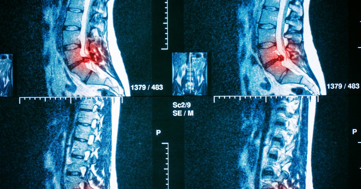
At NJBS, our clinicians frequently consult with patients who are highly anxious after reading an MRI report that lists multiple degenerative findings. It is a fundamental clinical truth that many notable imaging anomalies — such as mild disc bulges or arthritic facet joints — do not correlate to active physical pain and require no surgery whatsoever. An expert NJBS neurosurgeon treats your objective motor strength and dermatomal nerve pathways, not the static grey-scale markings on a radiology film.
A 52-year-old male presented with severe radiating leg pain caused by a lumbar disc herniation. Despite physical therapy, anti-inflammatory medications, and activity modification, his symptoms continued to interfere with work and daily activities.
MRI imaging confirmed compression of a single nerve root. However, flexion-extension X-rays demonstrated normal spinal alignment without evidence of spinal instability or abnormal vertebral movement.
Because the underlying issue was isolated nerve compression rather than structural instability, a minimally invasive decompression procedure was recommended. Following surgery, the patient’s leg pain improved significantly, and he returned to normal activities without requiring a spinal fusion.
A 59-year-old female sought evaluation for worsening lower back pain, leg pain, and difficulty walking. Imaging demonstrated lumbar spinal stenosis accompanied by spondylolisthesis — a condition in which one vertebra had shifted forward relative to the adjacent level.
While decompression was necessary to relieve pressure on the nerves, flexion-extension imaging demonstrated abnormal movement at the affected segment. Performing a decompression alone would have risked further destabilizing the spine.
Because both nerve compression and spinal instability were present, a decompression combined with spinal fusion was recommended. The procedure relieved the patient’s leg symptoms while also restoring long-term structural stability at the affected level.
Yes, there is a small statistical probability. If a patient undergoes a standalone decompression (such as a laminectomy) and the remaining disc or facet joints continue to degenerate normally over subsequent years, that specific motion segment can eventually develop mechanical instability. If this occurs and triggers recurrent nerve compression or severe mechanical back pain, a secondary revision surgery to execute a fusion may be considered.
Yes. While modern minimally invasive techniques have significantly reduced the surgical trauma of both procedures, a fusion inherently requires a longer overall recovery window. Standalone decompression patients are primarily waiting for local soft tissues and muscles to heal, which typically takes a few weeks. Fusion patients, however, must modify their activities for several months to protect the structural hardware while their body undergoes the biological process of growing new bone to fuse the vertebrae together.
If the structural instability is mild or low-grade, a highly specialized physical therapy protocol focused on deep core stabilization can be exceptionally effective. Strengthening the transverse abdominis, paraspinal, and oblique musculature creates a strong internal brace that can successfully minimize painful micro-movements. However, if the instability is severe, progressive, or causing objective neurological deficits like muscle wasting or foot drop, physical conditioning alone cannot override the mechanical failure of the bone structure.
For a standard single-level or two-level fusion, most patients report very little subjective loss in their global, day-to-day range of motion. This is because the remaining, unoperated joints above and below the fusion site naturally flex and adapt slightly more to compensate for the immobilized segment. A noticeable reduction in global flexibility typically only occurs when a patient requires an extensive, multi-level structural fusion spanning a large section of the spine.
Patients should always ask their specialist to walk them through their dynamic flexion/extension X-rays and structural scans. A qualified surgeon should be able to clearly point out objective metrics of mechanical instability, such as bone translation, facet joint destruction, or a structural spondylolisthesis. Seeking an independent second opinion from a board-certified neurosurgeon at NJBS is an excellent way to gain absolute clarity on whether a standalone decompression or a fusion is the safest path forward for your unique anatomy.
NJBS serves patients across northern New Jersey and the greater tri-state area, with offices in Paramus, Hackensack, Montclair, Montvale, Annandale, and Englewood. No referral is required to schedule a consultation.