Why MRI Findings Don’t Always Match Your Symptoms
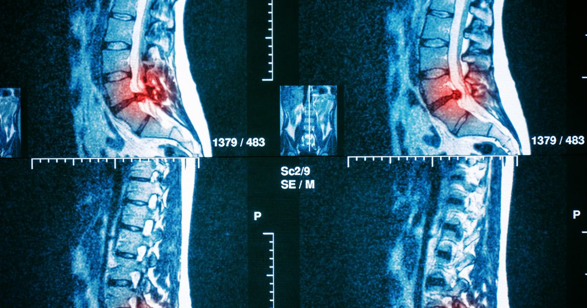
When a patient comes in holding a radiology report that lists disc herniations, stenosis, and degenerative changes at multiple spinal levels, the first question is rarely about anatomy. It is almost always some version of: “How bad is this? Is my spine falling apart? And why do I only hurt in one spot when the report makes it sound like there are problems everywhere?”
That disorientation is understandable. Terms like “degenerative changes at multiple levels” sound serious, and when a patient encounters them alongside real, ongoing pain, the assumption of a direct connection feels natural. But the relationship between what an MRI captures and what a patient actually experiences is far more nuanced than most radiology reports suggest.
An MRI is an extraordinary tool. It gives us structural detail that was invisible to medicine just a few decades ago. What it cannot do is measure how much a disc bulge is affecting your ability to put your shoes on in the morning, walk your dog, or sit through a workday. That information lives in you: in your history, your examination, and the story of how your symptoms have evolved. At New Jersey Brain and Spine, that is the information we weigh most heavily.
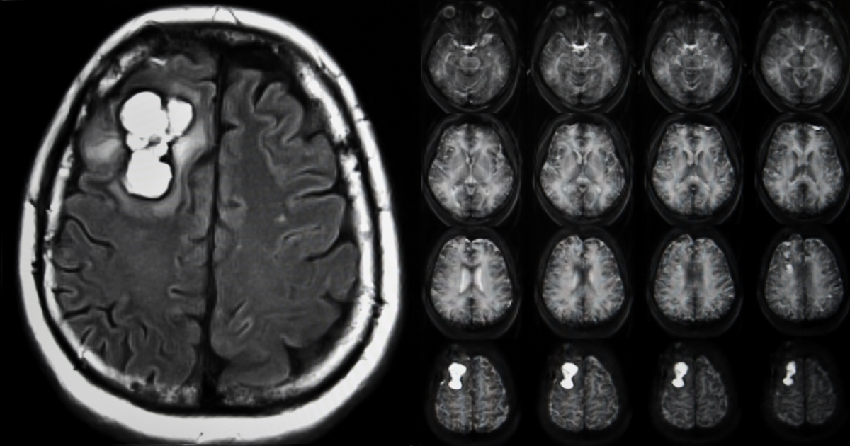
MRI, or magnetic resonance imaging, uses powerful magnetic fields and radio waves to produce detailed cross-sectional images of soft tissues, including discs, ligaments, nerves, and the spinal cord. It is static imaging: a snapshot of your spine at a single moment in time, while you are lying perfectly still.
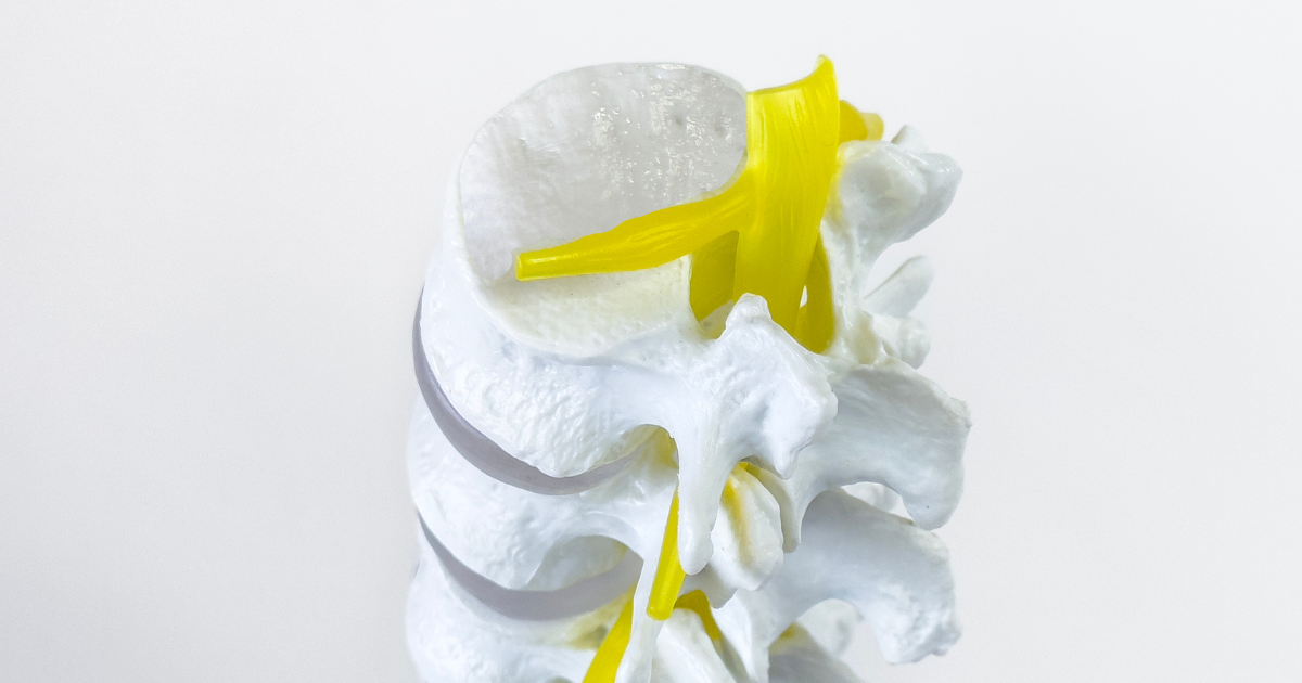
The spine is not a static structure. It is a dynamic system of interconnected moving segments. Each motion segment consists of two vertebrae, the disc between them, two facet joints behind them, and the ligaments and muscles that hold it all together. When you walk, bend, lift, or rotate, every segment participates. The symptoms you feel are generated in motion, under load, in real-world positions, not lying flat inside a scanner.
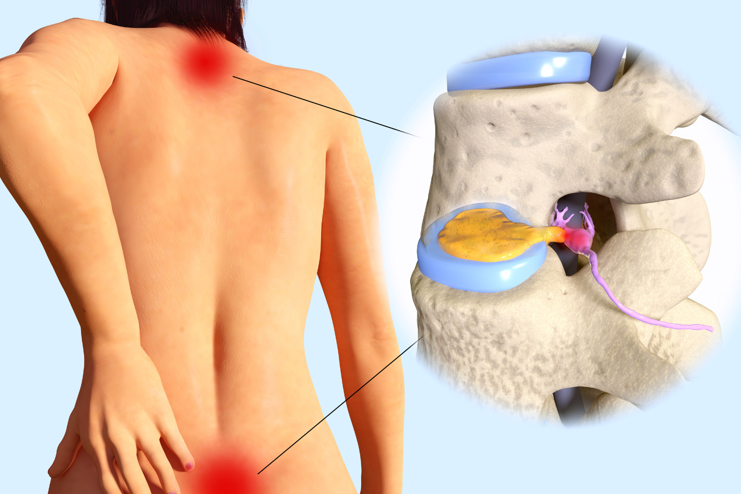
This creates a fundamental mismatch between what MRI shows and what symptoms mean. A disc can bulge without pressing on any nerve, producing no symptoms whatsoever. Degenerative changes on MRI are often a normal part of aging and may be entirely painless. Mild stenosis documented on imaging may correlate with severe walking limitation in one patient and no limitation in another. Asymptomatic findings, meaning abnormalities visible on scans in people who have no pain at all, are common across all age groups. And conversely, a patient with debilitating pain may have an MRI that appears relatively unremarkable.
MRI captures anatomy, not experience. A finding on a scan is a data point. It becomes clinically meaningful only when interpreted in the context of your specific symptoms, physical examination, and daily functional limitations.
This is not a clinical philosophy. It is a well-established pattern in the medical literature. Studies examining MRI scans in people with no back pain have found a consistently high prevalence of findings that would otherwise be labeled “abnormal.” The rates vary by age and by the specific finding, but the pattern holds: many structural changes visible on MRI are simply part of how spines age, not pathology requiring treatment.
Disc degeneration is found in roughly 37% of adults in their 20s, rising to over 90% by age 60. It is extremely common with age and does not reliably predict pain. Disc bulge is found in approximately 30% of asymptomatic adults under 40. A bulge alone, without nerve contact, often produces no symptoms. Disc herniation is identified in roughly 20 to 25% of pain-free adults. Many herniations resorb on their own and never cause symptoms. Facet joint degeneration is present in a large proportion of adults over 50. This is age-related arthritic change, common without symptoms. Mild spinal stenosis becomes increasingly common after age 50, and its functional impact varies enormously between individuals. Annular fissures, or disc tears, are found incidentally on many advanced MRI studies, and their significance depends on location, patient symptoms, and examination.
These numbers carry an important message: when your MRI report lists several findings, that list is not a verdict. It is a description of your anatomy at one point in time. At NJBS, we routinely see patients who were told their imaging looked “terrible” but who have minimal functional impairment, and others whose scans were read as “mild” but who are clearly experiencing significant nerve compromise. Neither scenario is unusual. Both require treating the person in front of us, not the image on the screen.
What matters, the part that requires clinical judgment rather than just a radiology read, is whether any of those findings actually explains your specific symptoms.
This patient was referred for evaluation after an MRI performed for mild intermittent low back pain showed multilevel degenerative disc disease, small disc bulges at three levels, and mild facet arthritis throughout the lumbar spine. Reading the report, she feared she would need surgery.
Her examination, however, told a different story. Her neurological function was entirely intact. Her pain was manageable and not progressive. Her daily activities, including her job, exercise routine, and household responsibilities, were largely unaffected. The imaging findings were consistent with her age and did not generate any nerve compression that correlated with a specific, localized symptom pattern.
Her care team explained that the findings described a common aging process, not a structural crisis. A structured physical therapy program focused on core stabilization and movement education produced excellent results. She left her appointment not with a surgical plan, but with a much clearer understanding of her spine and genuine confidence that she was not on the verge of structural collapse.
This patient presented with eight months of progressively worsening right leg pain radiating from his buttock to his foot, accompanied by numbness and tingling. His MRI was interpreted as showing only a “small” disc herniation at L4-L5 with “mild” nerve root contact. The radiologist’s language minimized what he was experiencing.
On careful clinical examination, however, he demonstrated clear weakness in his right foot and an absent ankle reflex, both signs of significant nerve root compromise. The “mild” imaging finding was, in his specific anatomy, causing meaningful nerve dysfunction. His care team coordinated targeted interventional treatment. When his symptoms failed to improve adequately after a thorough conservative trial, surgery was discussed as an appropriate next step. His outcome was excellent.
Note: These patient scenarios are representative, hypothetical examples used for illustrative purposes. Because every medical history is unique, actual treatment recommendations and surgical settings depend entirely on your individual health circumstances and a thorough evaluation by your care team.
Receiving a concerning MRI report does not automatically warrant a neurosurgical consultation. However, specialist evaluation is appropriate when your symptoms correlate with a specific finding, meaning your pain pattern, weakness, or numbness follows a nerve distribution that matches what imaging shows. It is also appropriate when conservative care has not improved your function after a reasonable period of 6 to 12 weeks, when your symptoms are progressing with worsening numbness, new weakness, or an expanding pain distribution, when new neurological findings appear on examination such as reflex changes or muscle weakness, or when your quality of daily life is significantly affected despite appropriate non-surgical management including physical therapy and interventional options such as epidural steroid injections.
Conversely, an incidental finding on an MRI in the absence of meaningful symptoms rarely requires specialist intervention. When in doubt, a conversation with a knowledgeable clinician who takes time to hear your full story, and not just review your imaging, is the right first step. New Jersey patients seeking a second opinion on a spine imaging report are always welcome at NJBS for exactly this kind of evaluation.
Cauda equina syndrome is a rare but serious condition that occurs when severe compression affects the bundle of nerve roots at the base of the spinal canal. It requires emergency treatment, and delay can result in permanent disability.
Call 911 or go to the nearest emergency room immediately if you experience saddle anesthesia, which is numbness or loss of sensation in the inner thighs, groin, buttocks, or genital area. Also seek emergency care for loss of bowel or bladder control, including sudden inability to urinate at all, bilateral leg weakness that is rapidly progressing in both legs simultaneously, or new sexual dysfunction that accompanies any of the symptoms above.
These symptoms represent a true neurological emergency. Do not wait for a scheduled appointment. Go to the emergency room immediately.
Degenerative changes on a spinal MRI are extremely common and, in many cases, represent a normal part of how the spine ages. The term sounds alarming because it contains the word “degeneration,” but it most often describes changes in disc hydration, disc height, or joint surfaces that occur over decades in most people, with or without pain. Whether these changes are clinically relevant depends entirely on whether they correspond to your specific symptoms and examination findings. A finding without a matching clinical picture rarely requires treatment.
In many patients, yes. Research consistently shows that disc herniations, even larger ones, frequently shrink or resorb over weeks to months as the body’s immune system processes the displaced disc material. This is one of the reasons conservative care is recommended first for most disc herniations: a meaningful percentage of patients improve without any intervention. The timeline and degree of natural resolution vary, but the potential for spontaneous improvement is real and well documented.
Back pain is one of the most complex symptoms in medicine, and structural findings on MRI represent only one possible contributor. Muscle imbalances, ligament strain, small joint irritation, movement pattern dysfunction, and central sensitization, a state in which the nervous system itself becomes heightened in its response to pain signals, can all produce significant pain without a clear structural correlate on imaging. A “normal” or near-normal MRI in someone with real pain does not mean the pain is not real. It means the cause may lie in something the MRI is not designed to capture.
An MRI finding changes the clinical picture most meaningfully when it directly correlates with your symptoms and examination. If your pain radiates in a specific nerve pattern and imaging shows compression of exactly that nerve, the finding is highly relevant. If imaging reveals significant instability, infection, tumor, or progressive cord compression, it will directly guide treatment decisions. The key question is not “what does the MRI show?” but “does what the MRI shows explain what you are experiencing?”
In most cases, surgery requires both a compelling clinical picture and an imaging finding that explains it. Surgery on a spine that looks structurally unremarkable carries a high risk of not helping, and potentially worsening things. That said, MRI is not infallible, and some findings are subtle or position-dependent. In select situations, additional studies such as dynamic X-rays, CT myelography, or provocative diagnostic injections can help clarify the relationship between anatomy and symptoms when standard MRI alone does not tell the full story.
NJBS serves patients across northern New Jersey and the greater tri-state area, with offices in Paramus, Hackensack, Montclair, Montvale, Annandale, and Englewood. No referral is required to schedule a consultation.