How Neurosurgeons Evaluate Nerve Damage
Patients come to us describing symptoms that do not fit neatly into the categories their previous workup has addressed. A hand that is progressively losing grip strength. Tingling that starts in the lower back and runs into the foot. Weakness in one arm that appeared without any clear injury. These presentations raise the same fundamental clinical question: is this a nerve problem, where in the nervous system is it originating, and what is actually happening to the nerve involved?
Evaluating nerve damage is one of the more nuanced aspects of neurosurgical practice. A scan shows structure, but it does not always tell us how your nerves are actually functioning. At New Jersey Brain and Spine, when we evaluate nerve damage, we look at your symptoms, your strength, your reflexes, and your daily life alongside any imaging. The goal of a neurosurgical nerve evaluation is to build a complete picture of what is happening and why, so that we can offer the most precise and effective path forward.
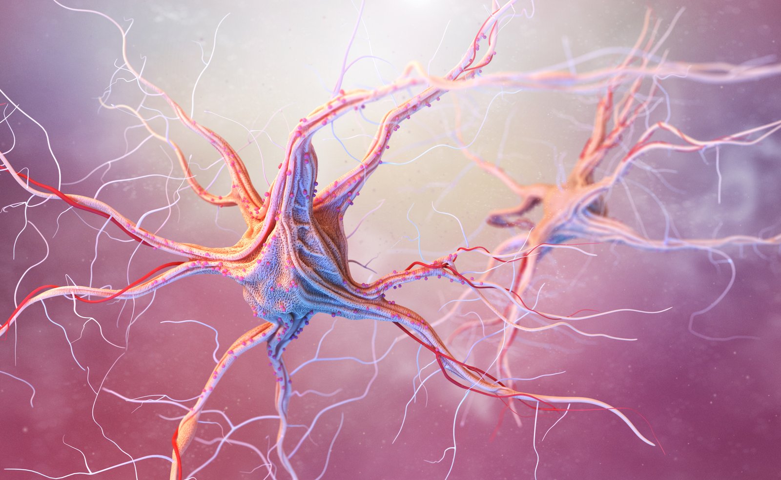
The nervous system is divided into the central nervous system, comprising the brain and spinal cord, and the peripheral nervous system, which includes the nerves that branch outward from the spinal cord to reach the limbs, organs, and skin. Damage can occur at any point along this pathway, and the location of the injury determines both the pattern of symptoms and the approach to treatment.
Nerves transmit signals in two directions. Motor signals travel from the brain and spinal cord outward to muscles, telling them to contract and produce movement. Sensory signals travel in the opposite direction, carrying information about touch, pain, temperature, and position from the body back to the brain. When a nerve is compressed, stretched, or otherwise injured, one or both of these signal pathways can be disrupted.
The degree of nerve injury exists on a spectrum. Mild compression may produce intermittent numbness or tingling that resolves when pressure is relieved. More sustained compression leads to persistent sensory changes and, over time, motor weakness. Severe or prolonged nerve injury can result in loss of muscle bulk and permanent functional deficits if not addressed. Understanding where on this spectrum a patient falls is central to determining the urgency and nature of intervention.
At NJBS, we also evaluate for signs of central versus peripheral nerve involvement, since the treatment approach differs significantly. A pinched nerve root in the spine (radiculopathy) is managed very differently from peripheral nerve entrapment at the wrist (carpal tunnel syndrome) or from spinal cord compression (myelopathy), even though patients with all three conditions may describe numbness in the hand.
A thorough nerve evaluation at NJBS begins with a detailed history. The pattern of onset, whether symptoms began suddenly or gradually, the distribution of numbness or weakness, any aggravating or relieving positions, and the impact on daily activities all provide diagnostic information that no scan can replace.
Physical examination focuses on several key areas. Muscle strength testing across specific muscle groups allows us to identify patterns of weakness that localize to specific nerve roots or peripheral nerves. Sensory testing maps the distribution of altered sensation against known dermatomal and nerve territory patterns. Reflex testing reveals the integrity of the reflex arc, which can pinpoint the spinal level or peripheral nerve involved. Coordination and gait assessment helps identify signs of myelopathy or central nervous system involvement.
Electrodiagnostic studies, including electromyography (EMG) and nerve conduction studies (NCS), are often used to objectively quantify nerve and muscle function. These tests measure the speed and amplitude of electrical signals traveling through specific nerves and assess whether individual muscles are receiving normal innervation. At NJBS, we use these studies selectively when the clinical picture requires additional objective data, particularly when the examination findings and imaging do not fully align.
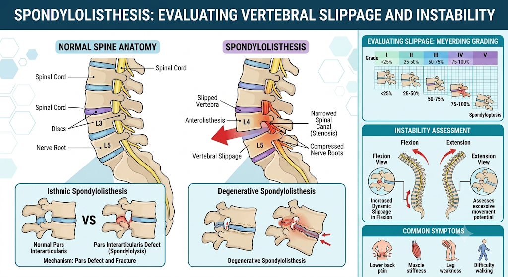
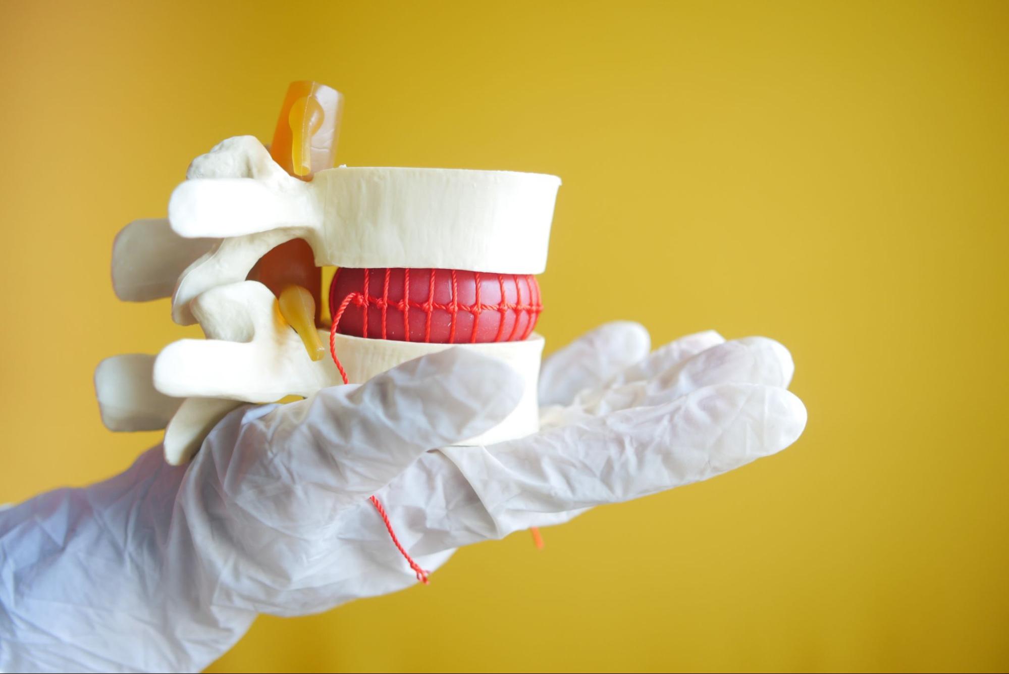
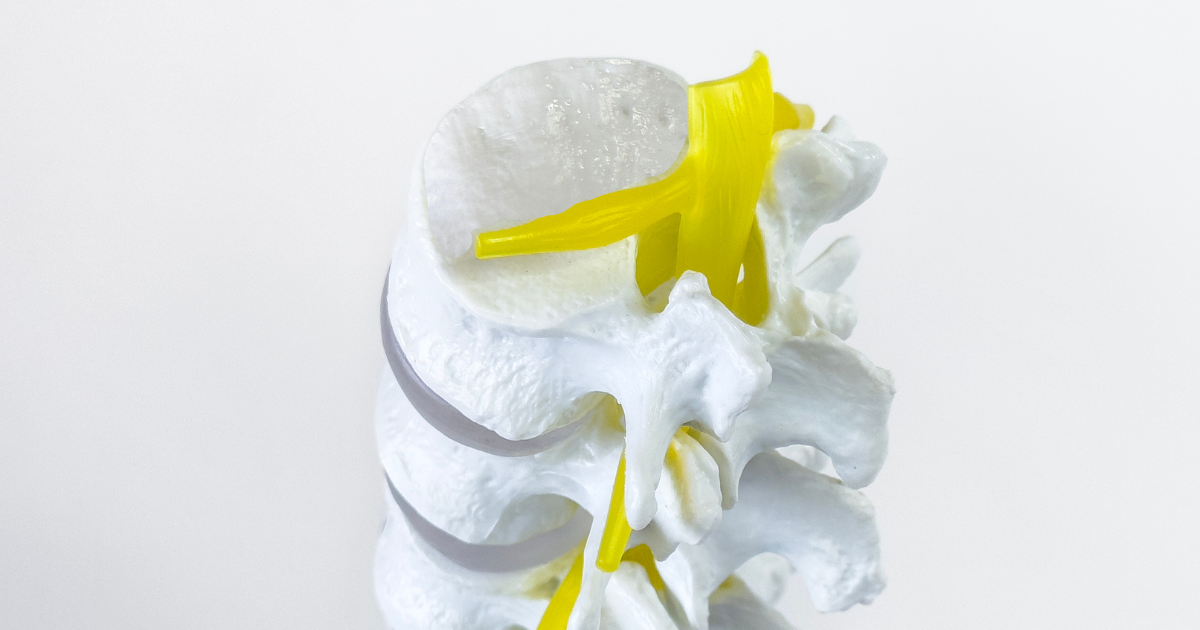
Radiculopathy occurs when a spinal nerve root is compressed or irritated, most commonly by a herniated disc or bone spur. Symptoms follow a specific dermatomal pattern corresponding to the affected root level: L4 radiculopathy produces pain and numbness into the shin, L5 into the top of the foot, and S1 into the heel and outer foot. Motor weakness follows the myotomal distribution of that root. Conservative care with physical therapy and, when indicated, epidural steroid injections resolves most cases of radiculopathy without surgery.
Myelopathy refers to dysfunction of the spinal cord itself, most commonly from cervical or thoracic stenosis compressing the cord directly. Unlike radiculopathy, myelopathy produces findings below the level of compression rather than in a single nerve root distribution. Patients may describe difficulty with fine motor tasks, changes in gait or balance, generalized leg weakness, or, in advanced cases, bowel and bladder dysfunction. Myelopathy is a condition that warrants careful evaluation and, in many cases, surgical decompression, as prolonged cord compression can lead to irreversible deficit.
Peripheral neuropathy involves damage to the peripheral nerves themselves, outside the spine. It can be caused by metabolic conditions such as diabetes, toxic exposures, inflammatory processes, or entrapment at specific anatomical sites. The treatment depends on identifying the underlying cause. Peripheral nerve entrapment, such as carpal tunnel syndrome or cubital tunnel syndrome, is often amenable to surgical decompression when conservative measures have not provided adequate relief.
At NJBS, a patient presenting with hand numbness and weakness receives an evaluation designed to distinguish between cervical myelopathy, cervical radiculopathy, and peripheral nerve entrapment before any treatment is recommended, because the appropriate intervention differs substantially depending on the correct diagnosis.
This patient presented with a several-month history of dropping objects, difficulty buttoning her shirt, and a sense that her hands felt less coordinated than they used to. She had intermittent neck stiffness but no significant neck pain or radicular arm symptoms.
Examination revealed mildly decreased grip strength bilaterally, hyperreflexia in the upper extremities, an abnormal Hoffmann sign bilaterally, and a broad-based gait. These findings were consistent with cervical myelopathy rather than a peripheral nerve problem. MRI confirmed significant cord compression at C5-C6 from a combination of disc herniation and ligamentous hypertrophy.
Given the progressive nature of her deficits and the degree of cord compression, surgical decompression was recommended. She underwent anterior cervical discectomy and fusion at C5-C6 with excellent postoperative recovery and stabilization of her hand function.
This patient developed acute right foot drop following a lifting injury at work. He could not dorsiflex his right foot and had numbness over the top of the foot and into the first web space. There was no back pain and no prior spine history.
Electrodiagnostic studies were ordered and confirmed an isolated peroneal nerve injury at the fibular head consistent with a compressive mechanism rather than an L4-L5 radiculopathy. MRI of the lumbar spine was unremarkable. Management focused on peroneal nerve decompression and physical therapy, avoiding an unnecessary spinal workup.
Note: These patient scenarios are representative, hypothetical examples used for illustrative purposes. Because every nerve injury and medical history is unique, actual treatment recommendations depend entirely on your individual circumstances and a thorough evaluation by your care team.
Nerve symptoms that are progressive, not improving with conservative measures, or affecting your ability to perform daily functions warrant specialist evaluation. Specific indications include new weakness in any limb, changes in coordination or balance, numbness or tingling that is expanding in distribution, and any change in bowel or bladder function. These symptoms should not be managed with a wait-and-see approach when they are worsening.
New Jersey patients experiencing neurological symptoms that have not been adequately explained or treated elsewhere are encouraged to seek evaluation at NJBS, where clinical examination and imaging are interpreted together to arrive at an accurate diagnosis before treatment is recommended.
Patients with lumbar spinal conditions should be aware of the symptoms of cauda equina syndrome, a surgical emergency caused by severe compression of the nerve roots at the base of the spinal canal. Call 911 or go to the nearest emergency room immediately if you develop saddle anesthesia (numbness in the inner thighs, groin, and genital region), loss of bowel or bladder control, sudden inability to urinate, or rapidly progressing bilateral leg weakness. Do not wait for a scheduled appointment. These symptoms require emergency evaluation.
The distribution of numbness provides the most important clue. Numbness that follows a dermatomal pattern down the arm or leg from the neck or back suggests a spinal origin such as radiculopathy. Numbness confined to the territory of a specific peripheral nerve, for example the thumb, index, and middle fingers in carpal tunnel syndrome, points to a peripheral entrapment. A careful clinical examination combined with electrodiagnostic testing and appropriate imaging can reliably distinguish between these sources in most cases.
Mild to moderate nerve compression, when the compressive source is removed, often produces meaningful recovery. Peripheral nerves have limited capacity to regenerate, at a rate of roughly one millimeter per day, which means that recovery from more severe injuries is slow and may be incomplete. Spinal cord injury does not follow the same regenerative capacity. This is why the timing of decompression matters: nerve deficits that have been present for a short time generally respond better to treatment than those that have been present for months or years.
Electromyography (EMG) measures the electrical activity of muscles at rest and during contraction. Combined with nerve conduction studies, it provides objective information about whether specific nerves are functioning normally and whether muscles are receiving adequate nerve signals. At NJBS, we order electrodiagnostic studies when the clinical examination and imaging do not clearly identify the source of nerve symptoms, when multiple potential diagnoses are being considered, or when objective baseline data is needed before initiating treatment.
No. MRI is excellent for identifying structural causes of nerve compression such as disc herniations, stenosis, and tumors. However, early or mild nerve injury may not produce visible changes on standard MRI sequences. Peripheral nerve conditions such as carpal tunnel syndrome are diagnosed primarily by examination and electrodiagnostic testing, not by imaging. A normal MRI does not exclude a clinically significant nerve problem.
NJBS serves patients across northern New Jersey and the greater tri-state area, with offices in Paramus, Hackensack, Montclair, Montvale, Annandale, and Englewood. No referral is required to schedule a consultation.